Review
. 2015 Aug 28;7(8):202-11.
doi: 10.4329/wjr.v7.i8.202.

Imaging of bone metastasis: An update

Affiliations
Review

Imaging of bone metastasis: An update

Gerard J O'Sullivan et al. World J Radiol. .

Abstract

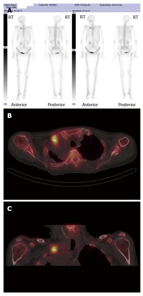
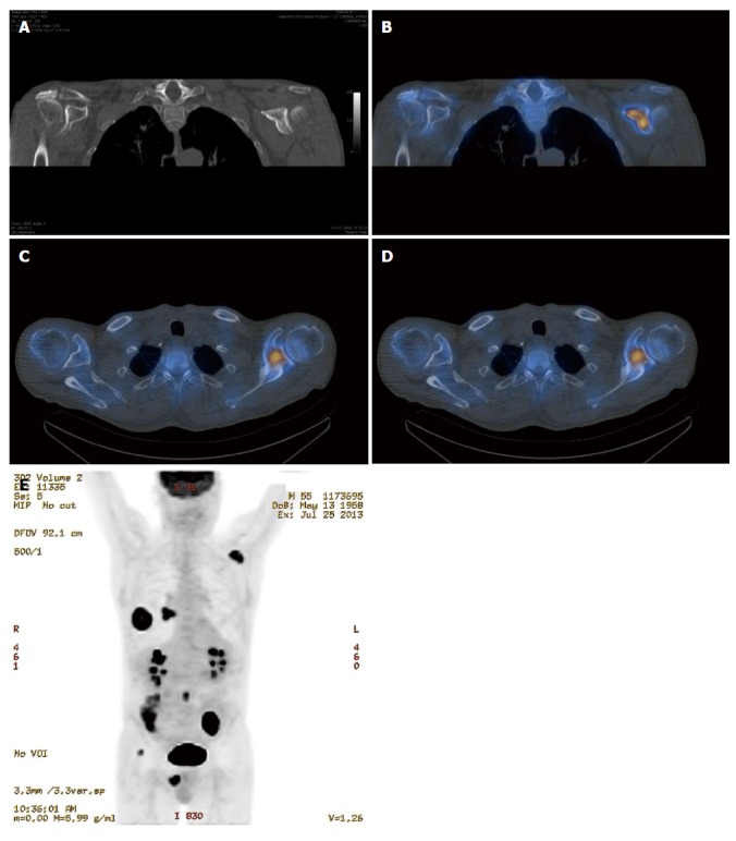
Early detection of skeletal metastasis is critical for accurate staging and optimal treatment. This paper briefly reviews our current understanding of the biological mechanisms through which tumours metastasise to bone and describes the available imaging methods to diagnose bone metastasis and monitor response to treatment. Among the various imaging modalities currently available for imaging skeletal metastasis, hybrid techniques which fuse morphological and functional data are the most sensitive and specific, and positron emission tomography (PET)/computed tomography and PET/magnetic resonance imaging will almost certainly continue to evolve and become increasingly important in this regard.

Keywords: Bone and bones; Computed tomography; Magnetic resonance imaging; Neoplasm metastasis; Radionuclide imaging.


Figures

Figure 1
Figure 1
Bone metastasis in the appendicular skeleton is most commonly due to primary lung malignancy. A: Axial computed tomography image of the upper thorax (soft tissue window) demonstrating a large right upper lobe mass with ipsilateral pulmonary and lymph node metastasis; B and C: PA and lateral views of the right thumb demonstrating a lytic metastatic deposit in the middle phalanx.
Figure 2
Figure 2
Magnetic resonance imaging is superior to plain radiography for detection of bone metastasis. A: Lateral lumbar spine radiograph demonstrates subtle sclerotic metastatic deposits at the inferior endplate of T12 and L1 from a primary breast malignancy; Sagittal T1 (B) and short tau inversion recovery (STIR) (C) images of the spine acquired one day later demonstrate diffuse bone metastasis (abnormal low T1 and high STIR signal in the bone marrow) which is not evident on the radiograph.
Figure 3
Figure 3
Diffuse bone metastasis on bone scintigraphy. Abnormal accumulation of radiotracer throughout the spine, most pronounced in the upper thoracic spine with additional pelvic and bilateral rib metastases in a patient with primary breast malignancy. Focal accumulation of radiotracer in the left antecubital fossa represents artefact at the radiotracer injection site.
Figure 4
Figure 4
Lytic bone metastases are poorly demonstrated on bone scintigraphy. Plain radiograph (A) demonstrating a lytic metastatic deposit in the right proximal humerus in a patient with a large right renal cell carcinoma (B); Corresponding abnormal low T1 and high short tau inversion recovery signal on magnetic resonance imaging (C and D); Only the small osteoblastic component of the metastatic deposit demonstrates abnormal accumulation of radiotracer on bone scintigraphy (E).
Figure 5
Figure 5
Single photon emission computed tomography has higher sensitivity and specificity for detection of bone metastasis when compared with bone scinitigraphy. A: Abnormal accumulation of radiotracer in the right clavicle on bone scintigraphy in a patient with primary lung malignancy; Axial (B) and coronal (C) single photon emission CT/CT images demonstrate the superior spatial and contrast resolution of this hybrid technique which enables improved detection and characterisation on bone metastases.
Figure 6
Figure 6
Single photon emission emission computed tomography-computed tomography is more sensitive for detection of bone metastasis than computed tomography alone. A: Coronal CT image of the left scapula (bone window) in a patient with primary lung malignancy does not demonstrate an aggressive bone lesion; Coronal and axial single photon emission CT/CT (B, C) and axial 18F fluorodeoxyglucose-positron emission tomography (FDG-PET)/CT (D) demonstrate abnormal radiotracer accumulation in the left clavicle consistent with bone metastasis; E: Coronal PET maximum intensity projection image demonstrating 18F FDG avid primary lung malignancy and right hilar lymph node metastasis in addition to the metastatic deposit in the left scapula.

References

    1. Bussard KM, Gay CV, Mastro AM. The bone microenvironment in metastasis; what is special about bone? Cancer Metastasis Rev. 2008;27:41–55. - PubMed
    1. Cuccurullo V, Cascini GL, Tamburrini O, Rotondo A, Mansi L. Bone metastases radiopharmaceuticals: an overview. Curr Radiopharm. 2013;6:41–47. - PubMed
    1. Vassiliou V, Andreopoulos D, Frangos S, Tselis N, Giannopoulou E, Lutz S. Bone metastases: assessment of therapeutic response through radiological and nuclear medicine imaging modalities. Clin Oncol (R Coll Radiol) 2011;23:632–645. - PubMed
    1. Yu HH, Tsai YY, Hoffe SE. Overview of diagnosis and management of metastatic disease to bone. Cancer Control. 2012;19:84–91. - PubMed
    1. Costelloe CM, Rohren EM, Madewell JE, Hamaoka T, Theriault RL, Yu TK, Lewis VO, Ma J, Stafford RJ, Tari AM, et al. Imaging bone metastases in breast cancer: techniques and recommendations for diagnosis. Lancet Oncol. 2009;10:606–614. - PubMed

LinkOut - more resources