. 2015 Sep 1;4(9):513-522.
doi: 10.1089/wound.2015.0638.

Angiogenic Potential of Cryopreserved Amniotic Membrane Is Enhanced Through Retention of All Tissue Components in Their Native State

Affiliations

Angiogenic Potential of Cryopreserved Amniotic Membrane Is Enhanced Through Retention of All Tissue Components in Their Native State

Yi Duan-Arnold et al. Adv Wound Care (New Rochelle). .

Abstract

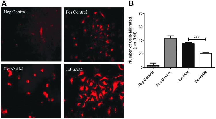
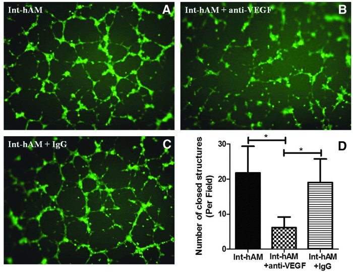
Objective: Chronic wounds have inadequate microvasculature (or blood vessels), resulting in poor healing. Both fresh human amniotic membrane (hAM) containing viable cells and devitalized hAM have been shown to stimulate angiogenesis in chronic wounds. However, the importance of retaining viable endogenous cells on the angiogenic activity of hAM remains unknown. To understand their role, we compared the angiogenic potential of intact cryopreserved hAM containing viable cells (int-hAM) with devitalized cryopreserved hAM (dev-hAM). Approach: The effects of conditioned medium (CM) derived from int-hAM and dev-hAM on endothelial cell migration and tube formation were compared. Int-hAM and dev-hAM CM and tissues were tested for key angiogenic factors, such as vascular endothelial growth factor (VEGF), basic fibroblast growth factor (bFGF), and platelet-derived growth factor-BB (PDGF-BB) after 7 days in culture. The role of VEGF in int-hAM-mediated tube formation was analyzed through inhibition of its activity by anti-VEGF antibody. Results: CM from int-hAM showed greater endothelial cell recruitment and tube formation compared with dev-hAM. Significantly higher levels of VEGF were detected in int-hAM CM after 1 week compared with dev-hAM CM. Int-hAM tissue also had significantly greater expression of VEGF and bFGF relative to dev-hAM. A similar trend was observed for PDGF-BB. Neutralization of VEGF in int-hAM CM significantly inhibited tube formation compared with int-hAM CM alone. Innovation and Conclusion: Preservation of all native hAM components, including viable endogenous cells, enhances the angiogenic effect of cryopreserved hAM. This effect is mediated through higher levels of angiogenic factors, especially VEGF, produced by int-hAM.


Figures

None
Yi Duan-Arnold, PhD
<b>Figure 1.</b>
Figure 1.
Hematoxilin & eosin (H&E) and collagen IV (Col IV) staining of human amniotic membranes (hAM). H&E staining of (A) fresh hAM, (B) viable intact cryopreserved hAM (int-hAM), and (C) devitalized cryopreserved hAM (dev-hAM). Brown colored stained Col IV in the basement membrane of (D) fresh-hAM, (E) int-hAM, and (F) dev-hAM. Scale bars: 50 μm (H&E) and 20 μm (Col IV).
<b>Figure 2.</b>
Figure 2.
Live and dead endogenous cell staining of hAM. Live and dead cells in epithelial and stromal layers of fresh hAM (A, B), int-hAM post-thaw (C, D), and dev-hAM post-thaw (E, F) were visualized microscopically using the LIVE/DEAD viability/cytotoxicity kit. Live cells stained green with calcein AM. Dead cells stained red with ethidium homodimer-1. Live and dead cells in int-hAM after 7 days in culture are shown in (G) and (H). All images were taken at 10×magnification.
<b>Figure 3.</b>
Figure 3.
Effect of hAM on human umbilical vein endothelial cells (HUVEC) migration. The migration of HUVEC induced by conditioned medium (CM) derived from int-hAM and dev-hAM was tested. HUVEC were incubated with CM overnight, and migrated HUVEC were stained with CM-DiI and visualized microscopically. The number of migrated HUVEC was counted, and results are presented as the number of cells migrated per microscopic field. Basal medium was used as a negative (Neg) control, and HUVEC growth medium was used as a positive (Pos) control. Representative images of migrated HUVEC at 10×magnification are shown in (A). Quantitative results are shown in (B). Data are presented as mean±standard deviation (SD) for one representative experiment out of three, with two biological replicates counted by three blinded operators. Student's t-test was used for statistical analysis. ***p<0.001.
<b>Figure 4.</b>
Figure 4.
Effect of hAM on HUVEC tube formation. Formation of closed, vessel-like structures by HUVEC was evaluated by culturing cells for 6 h with CM derived from int-hAM and dev-hAM. HUVEC were then labeled with calcein AM and visualized microscopically. The number of closed, tubular structures (an example of which is labeled with a white circle in Fig. 2A, positive control) formed by HUVEC was counted, and results are presented as the number of closed structures per microscopic field. Basal medium was used as a negative (Neg) control, and HUVEC growth medium was used as a positive (Pos) control. Representative images of HUVEC at 4×magnification are shown in (A). Results of quantitative analysis are shown in (B). Data are presented as mean±SD for one representative experiment out of three, with two biological replicates counted by three blinded operators. Student's t-test was used for statistical analysis. **p<0.01.
<b>Figure 5.</b>
Figure 5.
Levels of angiogenic growth factors in hAM after 7 days in culture. int-hAM and dev-hAM were cultured for 7 days. The CM and tissue lysates (TL) were collected and tested for vascular endothelial growth factor (VEGF) (A), basic fibroblast growth factor (bFGF) (B), and platelet-derived growth factor-BB (PDGF-BB) (C) by ELISA. The sum of growth factor content in CM and TL represents total values. Data are presented as mean±SD for one representative experiment out of three, with two biological replicates. Student's t-test was used for statistical analysis. *p<0.05, **p<0.01, and ***p<0.001.
<b>Figure 6.</b>
Figure 6.
Anti-VEGF neutralizing antibody blocked tube formation induced by CM derived from int-hAM. Int-hAM CM alone (A), or with 50 μg/mL anti-VEGF165 antibody (B), or with 50 μg/mL goat IgG control (C) were used in vitro tube formation assay. The number of closed, tubular structures was counted, and results are presented as the number of closed structures per microscopic field. Photographs of HUVEC were taken at 4×magnification. Results of quantitative analysis are shown in (D). Data are presented as mean±SD for one representative experiment out of two, with two biological replicates counted by three blinded operators. Student's t-test was used for statistical analysis. *p<0.05.

References

    1. Clark R. Overview and general considerations of wound repair. In: Clark RAF, Henson PM, eds. The Molecular and Cellular Biology of Wound Repair. Springer, Science & Business Media, New York; 1988:3–33
    1. Falanga V. Wound healing and its impairment in the diabetic foot. Lancet 2005;366:1736–1743 - PubMed
    1. Sen CK. Wound healing essentials: let there be oxygen. Wound Repair Regen 2009;17:1–18 - PMC - PubMed
    1. Ulrich D, Lichtenegger F, Unglaub F, Smeets R, Pallua N. Effect of chronic wound exudates and MMP-2/-9 inhibitor on angiogenesis in vitro. Plast Reconstruct Surg 2005;116:539–545 - PubMed
    1. Krisp C, Jacobsen F, McKay MJ, Molloy MP, Steinstraesser L, Wolters DA. Proteome analysis reveals antiangiogenic environments in chronic wounds of diabetes mellitus type 2 patients. Proteomics 2013;13:2670–2681 - PubMed