. 2015:2015:385402.
doi: 10.1155/2015/385402. Epub 2015 Aug 3.

Vaccination with an Attenuated Ferritin Mutant Protects Mice against Virulent Mycobacterium tuberculosis

Affiliations

Vaccination with an Attenuated Ferritin Mutant Protects Mice against Virulent Mycobacterium tuberculosis

Selvakumar Subbian et al. J Immunol Res. 2015.

Abstract

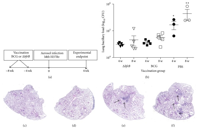
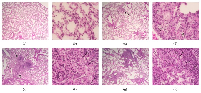
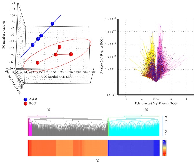
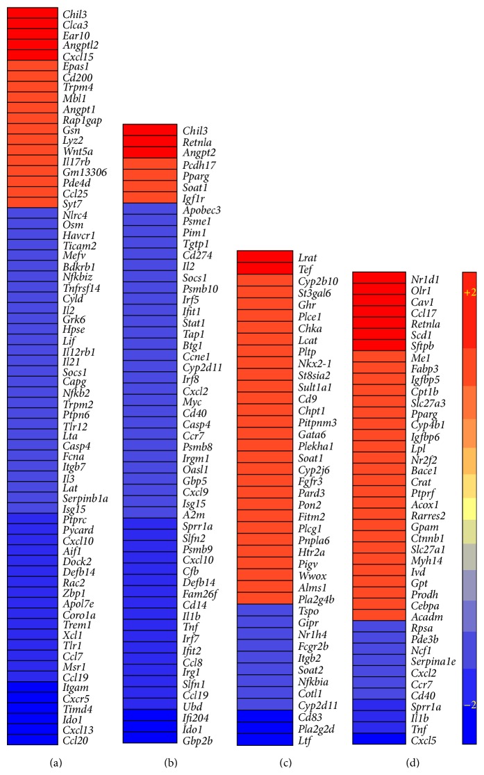
Mycobacterium tuberculosis the causative agent of tuberculosis affects millions of people worldwide. New tools for treatment and prevention of tuberculosis are urgently needed. We previously showed that a ferritin (bfrB) mutant of M. tuberculosis has altered iron homeostasis and increased sensitivity to antibiotics and to microbicidal effectors produced by activated macrophages. Most importantly, M. tuberculosis lacking BfrB is strongly attenuated in mice, especially, during the chronic phase of infection. In this study, we examined whether immunization with a bfrB mutant could confer protection against subsequent infection with virulent M. tuberculosis in a mouse model. The results show that the protection elicited by immunization with the bfrB mutant is comparable to BCG vaccination with respect to reduction of bacterial burden. However, significant distinctions in the disease pathology and host genome-wide lung transcriptome suggest improved containment of Mtb infection in animals vaccinated with the bfrB mutant, compared to BCG. We found that downmodulation of inflammatory response and enhanced fibrosis, compared to BCG vaccination, is associated with the protective response elicited by the bfrB mutant.


Figures

Figure 1
Figure 1
Experimental design, lung bacillary burden, and gross pathology of vaccinated and uninfected or Mtb-infected mice. (a) Schema of the mice vaccination schedule and Mtb infection experiment. (b) Lung bacillary load in mice infected with virulent Mtb H37Rv after vaccination with PBS (sham) or BCG or ΔbfrB for 4 ( P = 0.033) or 8 weeks (∗∗ P = 0.035). Gross lung pathology of vaccinated and uninfected or Mtb-infected-mice. (c) H&E stained lung section of BCG vaccinated (for 8 weeks) and uninfected mice. (d) H&E stained lung section of ΔbfrB vaccinated (for 8 weeks) and uninfected mice. (e) H&E stained lung section of BCG vaccinated (for 8 weeks) and Mtb-infected (for 4 weeks) mice. The arrows in (e) show a multifocal, coalescent granuloma. (f) H&E stained lung section of ΔbfrB vaccinated (for 8 weeks) and Mtb-infected (for 4 weeks) mice. The arrows in (f) show multiple, small granulomas.
Figure 2
Figure 2
Histopathology of vaccinated and uninfected or Mtb-infected mice lungs. ((a)-(b)) H&E stained lung section of BCG vaccinated (for 8 weeks) and uninfected mice. ((c)-(d)) H&E stained lung section of ΔbfrB vaccinated (for 8 weeks) and uninfected mice. The arrow in (c) shows cellular aggregation. The arrows in (d) show foamy histiocytes. ((e)-(f)) H&E stained lung section of BCG vaccinated (for 8 weeks) and Mtb-infected (for 4 weeks) mice. The arrows in (e) show a multifocal, coalescent granuloma. The arrows in (f) show lymphocyte cuff at the periphery of a granuloma. ((g)-(h)) H&E stained lung section of ΔbfrB vaccinated (for 8 weeks) and Mtb-infected (for 4 weeks) mice. The arrows in (g) show multiple, smaller granulomas (compared to (e)). The arrows in (h) show lymphocyte cuff at the periphery of a granuloma. Magnification: 4x ((a), (c), (e), and (g)) or 40x ((b), (d), (f), and (h)).
Figure 3
Figure 3
Genome-wide gene expression profiling of vaccinated and Mtb-infected mice lungs. (a) Principal component analysis of lung transcriptome data from ΔbfrB (blue) or BCG (red) vaccinated, Mtb-infected mice. The eclipse around each group denotes the standard deviation of the datasets. (b) Volcano plot of lung global gene expression showing the P value significance (y-axis; log scale) and fold change (x-axis). Upregulated genes are shaded in purple and downregulated genes are in yellow. N/C denotes no change. Each spot (blue) in the plot corresponds to a gene. (c) Intensity plot and dendrogram of significantly differentially expressed genes in the ΔbfrB vaccinated, compared to BCG vaccinated, Mtb-infected mice lungs. Upregulated genes are in red and downregulated genes are in blue. The color scale bar ranges from +3 (red) to −3 (blue).
Figure 4
Figure 4
Intensity plot of network genes differentially expressed in the vaccinated and Mtb-infected mice lungs. (a) Intensity plot of significantly differentially expressed genes involved in the inflammatory response network. (b) Intensity plot of significantly differentially expressed genes involved in the STAT-1 regulon network. (c) Intensity plot of significantly differentially expressed genes involved in the PC metabolism network. (d) Intensity plot of significantly differentially expressed genes involved in the PPAR-γ regulon network. The values plotted in (a)–(d) are different in fold change in the ΔbfrB vaccinated, compared to BCG vaccinated, Mtb-infected mice lungs. Upregulated genes are in red and downregulated genes are in blue. The color scale bar ranges from +2 (red) to −2 (blue).
Figure 5
Figure 5
Fibrosis in the lungs of vaccinated and uninfected or Mtb-infected mice. ((a)-(b)) Masson's trichrome stained lung section of BCG vaccinated (for 8 weeks) and uninfected (a) or Mtb-infected (b) mice. ((c)-(d)) Masson's trichrome stained lung section of ΔbfrB vaccinated (for 8 weeks) and uninfected (c) or Mtb-infected (d) mice. The arrows in (a) and (c) show basal level of fibrosis (blue color). The arrows in (b) show minimal fibrosis in the BCG vaccinated and Mtb-infected mice. The arrows in (d) show extensive fibrosis in the ΔbfrB vaccinated and Mtb-infected mice. Magnification: 4x ((a) and (c)) or 40x ((b) and (d)). (e) Intensity plot of significantly differentially expressed genes involved in the fibrosis network. The values plotted are the difference in fold change in the ΔbfrB vaccinated, compared to BCG vaccinated, Mtb-infected mice lungs. Upregulated genes are in red and downregulated genes are in blue. The color scale bar ranges from +2 (red) to −2 (blue).

References

    1. Rodriguez G. M. Control of iron metabolism in Mycobacterium tuberculosis . Trends in Microbiology. 2006;14(7):320–327. doi: 10.1016/j.tim.2006.05.006. - DOI - PubMed
    1. Prados-Rosales R., Weinrick B. C., Piqué D. G., Jacobs W. R., Jr., Casadevall A., Rodriguez G. M. Role for Mycobacterium tuberculosis membrane vesicles in iron acquisition. Journal of Bacteriology. 2014;196(6):1250–1256. doi: 10.1128/JB.01090-13. - DOI - PMC - PubMed
    1. Pandey R., Rodriguez G. M. A ferritin mutant of Mycobacterium tuberculosis is highly susceptible to killing by antibiotics and is unable to establish a chronic infection in mice. Infection and Immunity. 2012;80(10):3650–3659. doi: 10.1128/IAI.00229-12. - DOI - PMC - PubMed
    1. Koo M.-S., Subbian S., Kaplan G. Strain specific transcriptional response in Mycobacterium tuberculosis infected macrophages. Cell Communication and Signaling. 2012;10, article 2 doi: 10.1186/1478-811X-10-2. - DOI - PMC - PubMed
    1. Averbuch D., Chapgier A., Boisson-Dupuis S., Casanova J.-L., Engelhard D. The clinical spectrum of patients with deficiency of signal transducer and activator of transcription-1. Pediatric Infectious Disease Journal. 2011;30(4):352–355. doi: 10.1097/INF.0b013e3181fdff4a. - DOI - PubMed

Publication types

MeSH terms

LinkOut - more resources