. 2015 Oct 6;54(39):6029-37.
doi: 10.1021/acs.biochem.5b00622.

Dynamic Water-Mediated Hydrogen Bonding in a Collagen Model Peptide

Affiliations

Dynamic Water-Mediated Hydrogen Bonding in a Collagen Model Peptide

Iwen Fu et al. Biochemistry. .

Abstract

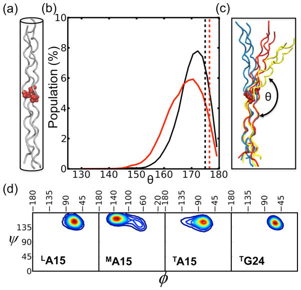
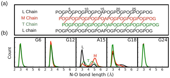
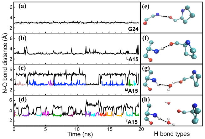
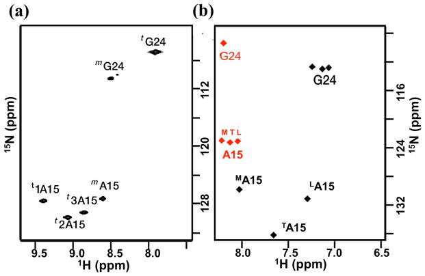
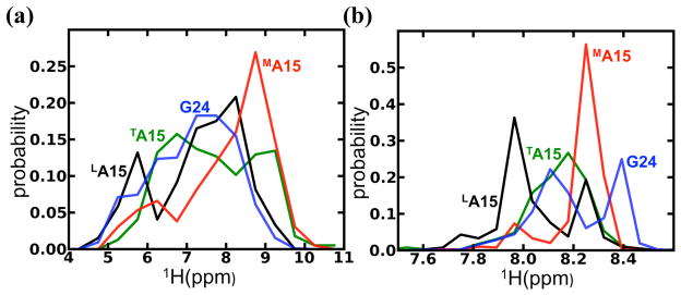
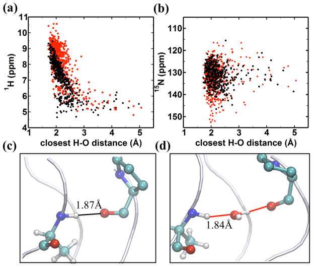
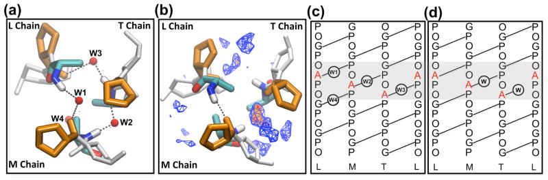
In the canonical (G-X-Y)(n) sequence of the fibrillar collagen triple helix, stabilizing direct interchain hydrogen bonding connects neighboring chains. Mutations of G can disrupt these interactions and are linked to connective tissue diseases. Here we integrate computational approaches with nuclear magnetic resonance (NMR) to obtain a dynamic view of hydrogen bonding distributions in the (POG)(4)(-)(POA)-(POG)(5) peptide, showing that the solution conformation, dynamics, and hydrogen bonding deviate from the reported X-ray crystal structure in many aspects. The simulations and NMR data provide clear evidence of inequivalent environments in the three chains. Molecular dynamics (MD) simulations indicate direct interchain hydrogen bonds in the leading chain, water bridges in the middle chain, and nonbridging waters in the trailing chain at the G → A substitution site. Theoretical calculations of NMR chemical shifts using a quantum fragmentation procedure can account for the unusual downfield NMR chemical shifts at the substitution sites and are used to assign the resonances to the individual chains. The NMR and MD data highlight the sensitivity of amide shifts to changes in the acceptor group from peptide carbonyls to water. The results are used to interpret solution NMR data for a variety of glycine substitutions and other sequence triplet interruptions to provide new connections between collagen sequences, their associated structures, dynamical behavior, and their ability to recognize collagen receptors.


Figures

Figure 1
Figure 1
(a) Schematic representation of the G→A crystal structure (PDB 1cag18) with Ala residues highlighted as red VDW balls; (b) the distributions of bend angles, θ (shown in (c). The dashed line indicates the angle of 175° and 177° calculated from crystal structure of (POG)10 (PDB 1v7h25) in black and G→A peptide in red, respectively. (c) MD selected snapshots corresponding to three bend angles (~180° in blue, ~160° in red, and ~145° in yellow) for the G→A peptide. (d) Ramachandran plot (ϕ,ψ) from the MD trajectories. Warmer colors represent higher populations.
Figure 2
Figure 2
Hydrogen bonding topology/patterns in the G→A peptide. (a) One residue staggering of the triple helix leads to leading (L), middle (M), and trailing (T) chains. (b) Distribution of N-O bond distances between N of the indicated residue and the carbonyl oxygen of the Pro in the neighboring chain. Chains L, M, and T are colored as black, red, and green, respectively.
Figure 3
Figure 3
Left panel: N-O bond distance between amide atom (N) of G24/A15 residues and the nearby oxygen atom (O) either from Pro carbonyl group (inter-chain distance colored as black) or a water molecule (solvent H-bond shown as other colors; water mediated H-bonds are always shown in blue and H-bonds to dangling water molecules are presented with different colors). For G24 in (a) and LA15 in (b), only inter-chain H-bonding is observed. For MA15 in (c) and TA15 in (d), alternate types of H bonds are found. Right panel shows the most representative H bond type corresponding to the residue in the left box and the H bond is indicated with a dashed line: (e) and (f) show inter-chain H bonds corresponding to G24 and LA15, respectively; (g) shows an interstitial water-mediated H bond for MA15 (colored in blue in panel c); (h) shows a dangling solvent H bond for TA15 (colored with many colors indicating different dangling solvent bonds in panel d).
Figure 4
Figure 4
(a) NMR HSQC spectrum23 of G→A peptide measured at 15°C, where three A15 trimers (t1A15, t2A15, and t3A15) are downfield shifted in the amide proton dimension relative to A15 monomer (mA15). (b) The AF-QMMM (black) and SHIFTX2 (red) calculations of chemical shifts on the 400 snapshots along 20-ns-long MD simulations of the G→A peptide in the trimer state.
Figure 5
Figure 5
Distribution of amide proton 1H chemical shifts using (a) AF-QMMM and (b) SHIFTX2 calculations on the 400 snapshots along a 20-ns-MD simulation. Note that the range of chemical shifts is significantly wider with the AF-QMMM calculations than with SHIFTX2 prediction. See Fig. S4 in Supporting Information for results using the SHIFTS program.
Figure 6
Figure 6
AF-QMMM chemical shift for the amide hydrogen in (a) and nitrogen in (b) of A15, demonstrating the dependence of chemical shift on the distance to the closest oxygen atom either from the carbonyl group (black dot) of a neighboring chain or from a water molecule (red dot) (c)&(d): representative snapshots with high downfield 1H chemical shifts attributed to H-bonding interactions shown by solid lines for (c) a direct H-bond with a 1H shift = 9.46 ppm for LA15; (d) for a water mediated H-bond with a 1H shift = 10.02 ppm for MA15.
Figure 7
Figure 7
Water distribution around the Gly substitution sites: (a) crystal structure18 and (b) MD solution from top view of the N terminal triple helix. In (a), W1 bridges the amide group of LA15 in the L chain and carbonyl group of Pro in the M chain. W2 and W3 are the water bridges of MA15 and TA15, respectively. W4 represents a water bridge for LG18. In (b), the MD simulated water density is shown with contour lines, which are coded by the total occupancy level of the volume. Volumes occupied > 75% are shown in red, > 50% in orange, and > 25% in blue. Crystal interstitial waters (W1–W3) are marked as red spheres; the three Ala are represented as licorice; Pro and Hyp are colored in orange and gray, respectively. Schematic of the hydrogen bonding topology around the Gly substitution sites (gray area) of a G→A peptide in the (c) crystal form and (d) solution form. Direct inter-chain hydrogen bonds are indicated as solid lines, hydrogen bonding to water is represented with the W label in the circle.

References

    1. Myllyharju J, Kivirikko KI. Collagens, Modifying enzymes and their mutations in humans, flies and worms. Trends Genet. 2004;20:33–43. - PubMed
    1. Shoulders MD, Raines RT. Collagen structure and stability. Annu Rev Biochem. 2009;78:929–958. - PMC - PubMed
    1. Heino J. The collagen family members as cell adhesion proteins. Bioessays. 2007;29:1001–1010. - PubMed
    1. Ramachandran GN, Kartha G. Structure of collagen. Nature. 1955;176:593–595. - PubMed
    1. Rich A, Crick FH. The structure of collagen. Nature. 1955;176:915–916. - PubMed

Publication types

LinkOut - more resources