

Development of the Synarcual in the Elephant Sharks (Holocephali; Chondrichthyes): Implications for Vertebral Formation and Fusion
- PMID: 26339918
- PMCID: PMC4560447
- DOI: 10.1371/journal.pone.0135138
Development of the Synarcual in the Elephant Sharks (Holocephali; Chondrichthyes): Implications for Vertebral Formation and Fusion
Abstract
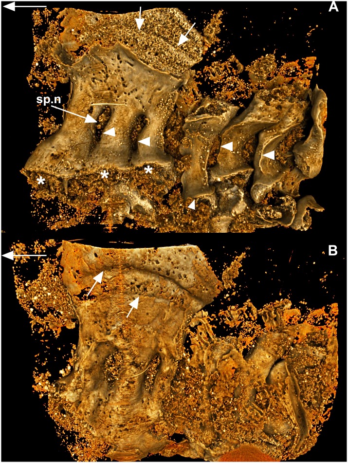
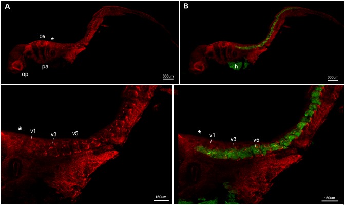
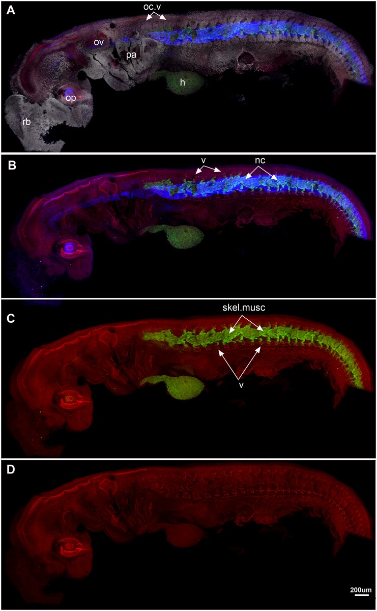
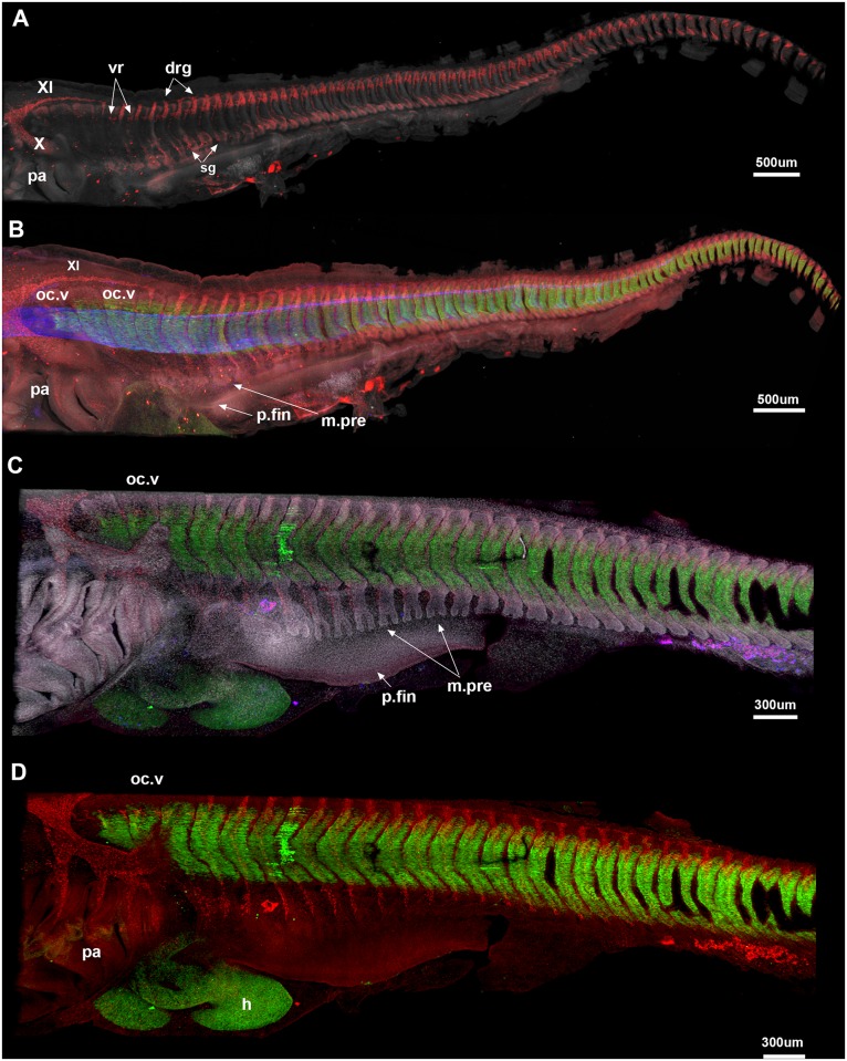
The synarcual is a structure incorporating multiple elements of two or more anterior vertebrae of the axial skeleton, forming immediately posterior to the cranium. It has been convergently acquired in the fossil group 'Placodermi', in Chondrichthyes (Holocephali, Batoidea), within the teleost group Syngnathiformes, and to varying degrees in a range of mammalian taxa. In addition, cervical vertebral fusion presents as an abnormal pathology in a variety of human disorders. Vertebrae develop from axially arranged somites, so that fusion could result from a failure of somite segmentation early in development, or from later heterotopic development of intervertebral bone or cartilage. Examination of early developmental stages indicates that in the Batoidea and the 'Placodermi', individual vertebrae developed normally and only later become incorporated into the synarcual, implying regular somite segmentation and vertebral development. Here we show that in the holocephalan Callorhinchus milii, uniform and regular vertebral segmentation also occurs, with anterior individual vertebra developing separately with subsequent fusion into a synarcual. Vertebral elements forming directly behind the synarcual continue to be incorporated into the synarcual through growth. This appears to be a common pattern through the Vertebrata. Research into human disorders, presenting as cervical fusion at birth, focuses on gene misexpression studies in humans and other mammals such as the mouse. However, in chondrichthyans, vertebral fusion represents the normal morphology, moreover, taxa such Leucoraja (Batoidea) and Callorhinchus (Holocephali) are increasingly used as laboratory animals, and the Callorhinchus genome has been sequenced and is available for study. Our observations on synarcual development in three major groups of early jawed vertebrates indicate that fusion involves heterotopic cartilage and perichondral bone/mineralised cartilage developing outside the regular skeleton. We suggest that chondrichthyans have potential as ideal extant models for identifying the genes involved in these processes, for application to human skeletal heterotopic disorders.
Conflict of interest statement
Figures
Similar articles
-
Fusion, gene misexpression and homeotic transformations in vertebral development of the gnathostome stem group (Placodermi).Int J Dev Biol. 2010;54(1):71-80. doi: 10.1387/ijdb.072508zj. Int J Dev Biol. 2010. PMID: 19876847
-
Mineralization of the Callorhinchus Vertebral Column (Holocephali; Chondrichthyes).Front Genet. 2020 Nov 26;11:571694. doi: 10.3389/fgene.2020.571694. eCollection 2020. Front Genet. 2020. PMID: 33329708 Free PMC article.
-
The synarcual cartilage of batoids with emphasis on the synarcual of Rajidae.J Morphol. 2011 Dec;272(12):1444-63. doi: 10.1002/jmor.10996. Epub 2011 Jul 20. J Morphol. 2011. PMID: 21780157
-
Architectural and ultrastructural features of tessellated calcified cartilage in modern and extinct chondrichthyan fishes.J Fish Biol. 2021 Apr;98(4):919-941. doi: 10.1111/jfb.14376. Epub 2020 Jul 1. J Fish Biol. 2021. PMID: 32388865 Review.
-
Building the backbone: the development and evolution of vertebral patterning.Development. 2015 May 15;142(10):1733-44. doi: 10.1242/dev.118950. Development. 2015. PMID: 25968309 Review.
Cited by
-
The Natural History Museum Data Portal.Database (Oxford). 2019 Jan 1;2019:baz038. doi: 10.1093/database/baz038. Database (Oxford). 2019. PMID: 30985890 Free PMC article.
-
Endoskeletal mineralization in chimaera and a comparative guide to tessellated cartilage in chondrichthyan fishes (sharks, rays and chimaera).J R Soc Interface. 2020 Oct;17(171):20200474. doi: 10.1098/rsif.2020.0474. Epub 2020 Oct 14. J R Soc Interface. 2020. PMID: 33050779 Free PMC article.
-
Phenotypic regionalization of the vertebral column in the thorny skate Amblyraja radiata: Stability and variation.J Anat. 2022 Feb;240(2):253-267. doi: 10.1111/joa.13551. Epub 2021 Sep 19. J Anat. 2022. PMID: 34542171 Free PMC article.
-
Mineralized Cartilage and Bone-Like Tissues in Chondrichthyans Offer Potential Insights Into the Evolution and Development of Mineralized Tissues in the Vertebrate Endoskeleton.Front Genet. 2021 Dec 22;12:762042. doi: 10.3389/fgene.2021.762042. eCollection 2021. Front Genet. 2021. PMID: 35003210 Free PMC article. Review.
-
Migratory patterns and evolutionary plasticity of cranial neural crest cells in ray-finned fishes.Dev Biol. 2020 Nov 1;467(1-2):14-29. doi: 10.1016/j.ydbio.2020.08.007. Epub 2020 Aug 21. Dev Biol. 2020. PMID: 32835652 Free PMC article.
References
-
- Gadow H. The evolution of the vertebral column: a contribution to the study of the vertebrate phylogeny. 1933; Cambridge: Cambridge University Press.
-
- Christ B, Schmidt C, Huang R, Wilting J, Brand-Saberi B. Segmentation of the vertebrate body. Anat Embryol. 1998;197: 1–8. - PubMed
-
- Monsoro-Burq AH. Sclerotome development and morphogenesis: when experimental embryology meets genetics. Int J Dev Biol. 2005;49: 301–308. - PubMed
-
- Arratia G, Schultze HP, Casciotta J. Vertebral column and associated elements in dipnoans and comparison with other fishes: development and homology. J Morphol. 2001;250: 101–72. - PubMed
-
- Aulehla A, Herrmann BG. Segmentation in vertebrates: clock and gradient finally joined. Gene Dev. 2004;18: 2060–2067. - PubMed
Publication types
MeSH terms
LinkOut - more resources
Full Text Sources
Other Literature Sources
