. 2015 Sep 4;10(9):e0137468.
doi: 10.1371/journal.pone.0137468. eCollection 2015.

The Expression of a Novel Mitochondrially-Encoded Gene in Gonadic Precursors May Drive Paternal Inheritance of Mitochondria

Affiliations

The Expression of a Novel Mitochondrially-Encoded Gene in Gonadic Precursors May Drive Paternal Inheritance of Mitochondria

Liliana Milani et al. PLoS One. .

Abstract

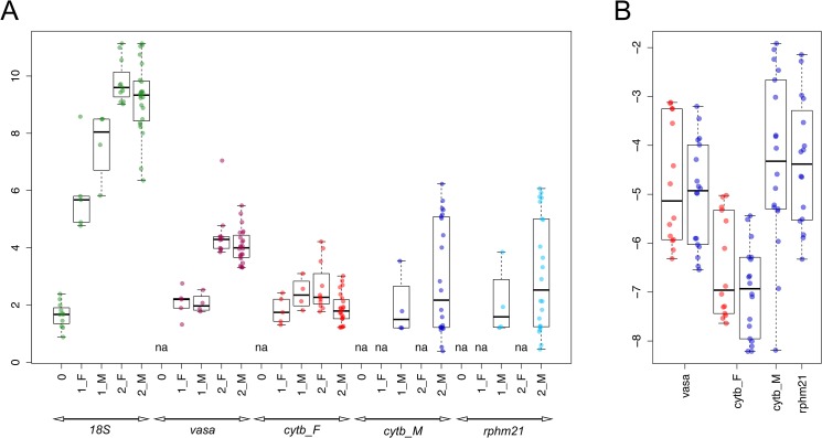
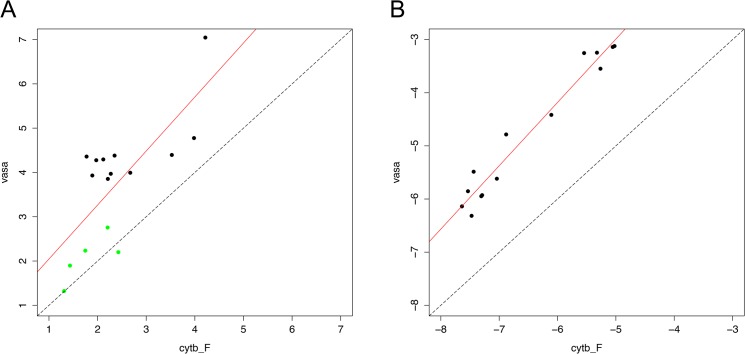
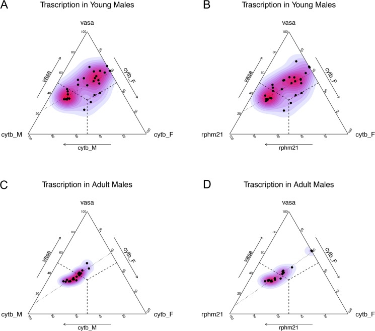
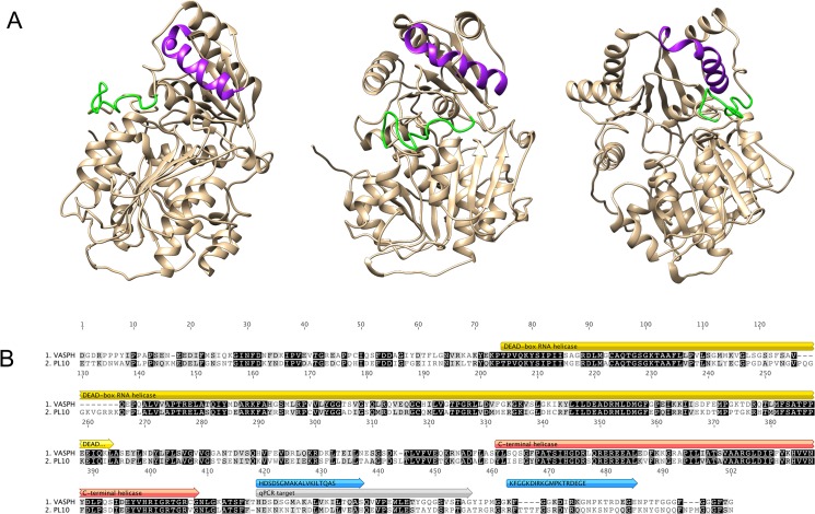
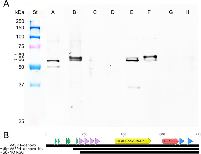
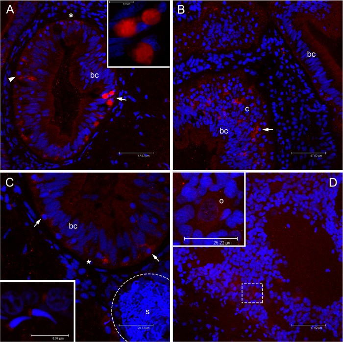
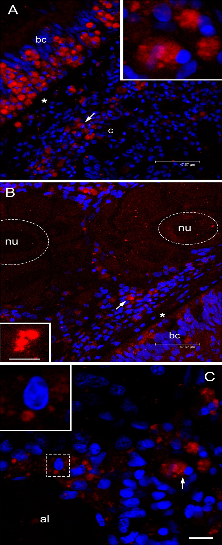
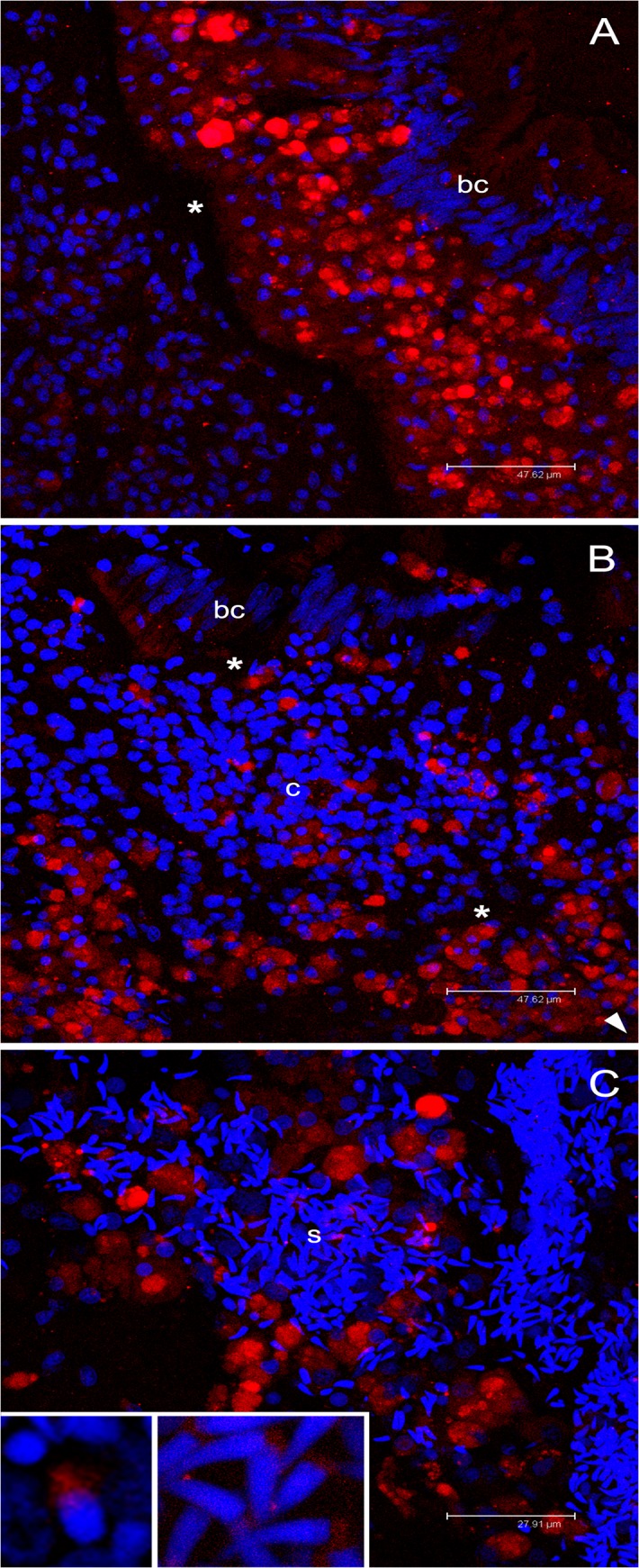
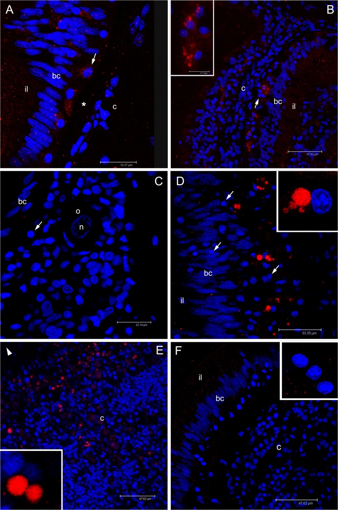
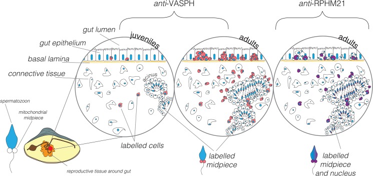
Mitochondria have an active role in germ line development, and their inheritance dynamics are relevant to this process. Recently, a novel protein (RPHM21) was shown to be encoded in sperm by the male-transmitted mtDNA of Ruditapes philippinarum, a species with Doubly Uniparental Inheritance (DUI) of mitochondria. In silico analyses suggested a viral origin of RPHM21, and we hypothesized that the endogenization of a viral element provided sperm mitochondria of R. philippinarum with the ability to invade male germ line, thus being transmitted to the progeny. In this work we investigated the dynamics of germ line development in relation to mitochondrial transcription and expression patterns using qPCR and specific antibodies targeting the germ line marker VASPH (R. philippinarum VASA homolog), and RPHM21. Based on the experimental results we conclude that both targets are localized in the primordial germ cells (PGCs) of males, but while VASPH is detected in all PGCs, RPHM21 appears to be expressed only in a subpopulation of them. Since it has been predicted that RPHM21 might have a role in cell proliferation and migration, we here suggest that PGCs expressing it might gain advantage over others and undertake spermatogenesis, accounting for RPHM21 presence in all spermatozoa. Understanding how foreign sequence endogenization and co-option can modify the biology of an organism is of particular importance to assess the impact of such events on evolution.


Conflict of interest statement

Competing Interests: The authors have declared that no competing interests exist.

Figures

Fig 1
Fig 1. Transcription levels in juveniles (A) and adults (B).
(A) 0 = class B0; 1 = class B1; 2 = class B2; F = Females; M = Males. y axis: Log10 copy number ("absolute quantification" with standard dilutions). (B) Red = Females; blue = Males. y axis: Log10 copy number relative to 18S (relative quantification with 18S as nuclear endogenous control).
Fig 2
Fig 2. Transcription level correlations in juvenile (A) and adult (B) females.
In females, vasph transcription resulted to be strongly correlated to mitochondrial transcription (statistics in S4 Table). Axes in A: Log10 copy number ("absolute quantification" with standard dilutions). Axes in B: Log10 copy number relative to 18S (relative quantification with 18S as nuclear endogenous control). In (A): green dots = B1 class; black dots = B2 class.
Fig 3
Fig 3. Transcription level in juvenile (A, B) and adult (C, D) males.
(A) Transcription of vasph, cytb_M and cytb_F in young males. (B) Transcription of vasph, rphm21 and cytb_F in young males. (C) Transcription of vasph, cytb_M and cytb_F in adult males. (D) Transcription of vasph, rphm21 and cytb_F in adult males. Color gradient: kernel density estimation of the distribution. Dashed lines: middle segments representing equal transcription of the targets indicated at the vertexes of the orthogonal side. The three middle segments meet in the barycenter (i.e. the point of equal transcription of the three targets). Dotted lines: smoothed conditional mean of the distribution.
Fig 4
Fig 4. VASA homolog of Ruditapes philippinarum (VASPH).
(A) VASPH structure model. Chimera 1.8.1 was used to model the protein structure. Peptides used for antibody production are highlighted: HDS in purple; KFG in green. (B) Alignment of VASPH and PL10 of R. philippinarum. Peptide location (blue), qPCR target (grey) and protein main domains (yellow and red) are highlighted.
Fig 5
Fig 5. Detection of VASPH variants.
(A) Anti-VASPH specificity: Western blots of ovary extracts (Oe) and testis extracts (Te) of adults. From left to right: St: protein standard. A: Oe/anti-VASPH-HDS. B: Te/anti-VASPH-HDS. C: Oe/anti-VASPH-HDS control. D: Te/anti-VASPH-HDS control. E: Oe/anti-VASPH-KFG. F: Te/anti-VASPH-KFG. G: Oe/anti-VASPH-KFG control. H: Te/anti-VASPH-KFG control. Western blots were obtained with the loading of 15 μg of homogenate per lane. (B) Different VASPH transcript assemblies highlighted different N-terminus of the protein. RGG motifs (green), zinc fingers (violet), protein main domains (yellow and red), and peptide location (blue) are highlighted.
Fig 6
Fig 6. Immunolocalization of VASPH in germ cells of juveniles approaching their first reproductive season.
(A) In juvenile clams, anti-VASPH highlighted many immunospots in germ cells with rounded nucleus (PGCs) localized in the thickness of intestinal epithelium, between unstained batiprismatic cells (bc) of the gut, and the basal lamina (asterisk). In some germ cells, the immunospots appear separated (arrowhead), while in other cells the spots aggregate at a side of the cell cytoplasm (arrow; magnification in the inset). Scale bar = 47.62 μm (inset scale bar = 8.61 μm). (B) Stained germ cells (arrow) are also visible in the connective tissue (c) between two intestinal loops. Scale bar = 47.62 μm. (C) Portion of young male section that shows an acinus (dashed circle) full of sperm (s) close to an intestinal loop in which some stained germ cells are present (arrows). The inset shows a magnification of two spermatozoa with a lightly stained mitochondrial midpiece and a spermatid (up right) with a big immunostained spot near the nucleus. Scale bar = 26.12 μm (inset scale bar = 8.07 μm). (D) Portion of young female section that shows early oocytes at different stage of development with a light VASPH staining in the cytoplasm. In the inset, a magnification of an early oocyte (o) showing few small granules. Scale bar = 47.62 μm (inset scale bar = 25.22 μm). Red: VASPH staining; blue: nuclear staining.
Fig 7
Fig 7. Immunolocalization of VASPH in germ cells of gametogenic adult females.
(A) Section with a portion of intestine and connective tissue. Inside the intestinal epithelium, among batiprismatic cells (bc), a strong proliferation of VASPH stained germ cells (arrow) is observed. Many germ cells (arrow; magnified in the inset) have passed the basal lamina (asterisk) to the connective tissue (c). Scale bar = 47.57 μm. (B) In the connective tissue, in proximity of the intestine, germ cells (arrow) surround acini full of eggs (two eggs are highlighted with a dashed oval; nu: nucleus). In the egg, small stained granules are scattered in the cytoplasm (inset: granule magnification). Scale bar = 47.62 μm (inset scale bar = 4.87 μm). (C) At the periphery of an acinus lumen (al), very early oocytes of about 10 μm show big stained spots (one oocyte is magnified in the inset). Scale bar = 10.54 μm. Red: VASPH staining; blue: nuclear staining.
Fig 8
Fig 8. Immunolocalization of VASPH in germ cells of gametogenic adult males.
(A) Strong proliferation of VASPH-stained germ cells in the intestinal epithelium at one side of the basal lamina (asterisk). Batiprismatic cells (bc) resulted VASPH-unstained. Scale bar = 47.62 μm. (B) Many stained germ cells in the connective tissue (c) between two intestinal loops (the arrowhead points to the position of other batiprismatic cells of the gut). Scale bar = 47.62 μm. (C) High magnification of a portion of male acinus showing many spermatozoa that fill the lumen. Inset on the left: spermatid with VASPH staining limited at the posterior part of the elongating nucleus. Inset on the right: several spermatozoa showing an even more reduced labelling in the mitochondrial midpiece. Scale bar = 27.91 μm. Red: VASPH staining; blue: nuclear staining.
Fig 9
Fig 9. Immunolocalization of RPHM21 in germ cells.
(A) In juveniles, the immunological reaction highlighted some rounded-nucleus cells (germ cells) with a diffused cytoplasmic RPHM21 staining (arrow) between unstained batiprismatic cells (bc) and the basal lamina (asterisk). Intestinal lumen = il; connective tissue = c. Scale bar = 18.37 μm. (B) In some sections of juvenile animals, germ cells are visible in the connective tissue (c) and show big stained spots (inset). Scale bar = 47.62 μm (inset scale bar = 11.11 μm). (C) In some female sections, simple acini, at the beginning of their organization, sometimes containing a single oocyte (o), were found. In these female sections, germ cells were visible (arrow) but no RPHM21 staining was present. n = oocyte nucleus. Scale bar = 22.14 μm. (D) Adult male section that shows RPHM21 stained germ cells close to batiprismatic cells (bc); some germ cells do not show any RPHM21 staining (arrow). Scale bar = 33.35 μm. (E) In male connective tissue, several RPHM21-stained germ cells are found (the arrowhead points to intestine position). The inset shows magnified RPHM21stained germ cells. Scale bar = 47.62 μm. (F) In adult female sections, no RPHM21-staining was detected in germ cells (magnified in the inset). Scale bar = 47.62 μm. Red: RPHM21 staining; blue: nuclear staining.
Fig 10
Fig 10. Scheme of RPHM21 and VASPH immunolocalization in germ cells during male gonad formation.
VASPH expression (in red): in juvenile males (first circle on the left) few, stained PGCs are localized in the intestine among batiprismatic cells, and other stained germ cells are found in the connective tissue or around the few simple-structured acini localized in the connective tissue. In gametogenic males (circle in the middle) PGCs are massively proliferating among batiprismatic cells and are strongly immunostained. In mature male acini full of spermatozoa, a diffused VASPH-staining is present in the spermatogenic cells located near the acinus wall (see also [10]). Spermatozoon midpiece appears slightly stained. RPHM21 expression (in violet): only a subpopulation of PGCs located in the intestinal epithelium appears to express RPHM21, other PGCs, recognizable for their round nucleus, result completely negative to the RPHM21 staining. Some cells with a weak RPHM21 labelling (spermatogenic cells) are recognizable in the acinus wall [41]. RPHM21 is expressed in mature spermatozoa localized in the acinus lumen, both in mitochondria and the nucleus [41]. The staining of both factors (VASPH and RPHM21) is almost always condensed in a big cluster at one side of the cell cytoplasm.

Similar articles

Cited by

References

    1. Extavour CG, Akam M. Mechanisms of germ cell specification across the metazoans: epigenesis and preformation. Development. 2003; 130: 5869ng (sp - PubMed
    1. Matova N, Cooley L. Comparative aspects of animal oogenesis. Dev Biol. 2001; 231: 291–320. - PubMed
    1. Kloc M, Bilinski S, Etkin LD. The Balbiani body and germ cell determinants: 150 years later. Curr Top Dev Biol. 2004; 59: 12003 - PubMed
    1. Reunov A. Structures related to the germ plasm in mouse. Zygote. 2006; 14: 231–238. - PubMed
    1. Parvinen M. The chromatoid body in spermatogenesis. Int J Androl. 2005; 28: 189238.r - PubMed

Publication types

MeSH terms

Substances

LinkOut - more resources