. 2015 Sep 1;16(9):20859-72.
doi: 10.3390/ijms160920859.

Optimization and Evaluation of a Chitosan/Hydroxypropyl Methylcellulose Hydrogel Containing Toluidine Blue O for Antimicrobial Photodynamic Inactivation

Affiliations

Optimization and Evaluation of a Chitosan/Hydroxypropyl Methylcellulose Hydrogel Containing Toluidine Blue O for Antimicrobial Photodynamic Inactivation

Chueh-Pin Chen et al. Int J Mol Sci. .

Abstract

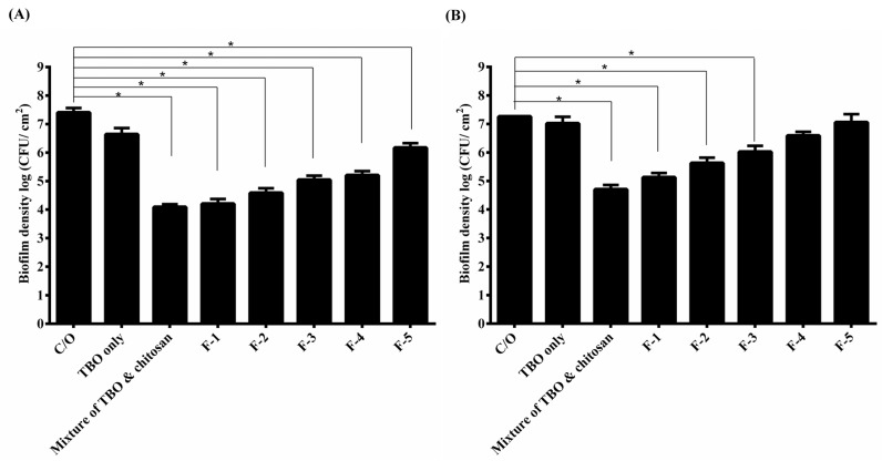
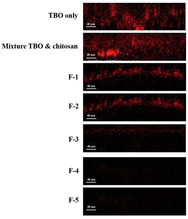
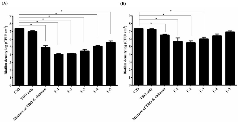
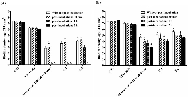
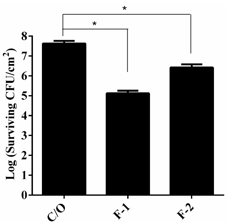
Photodynamic inactivation (PDI) combined with chitosan has been shown as a promising antimicrobial approach. The purpose of this study was to develop a chitosan hydrogel containing hydroxypropyl methylcellulose (HPMC), chitosan and toluidine blue O (TBO) to improve the bactericidal efficacy for topical application in clinics. The PDI efficacy of hydrogel was examined in vitro against the biofilms of Staphylococcus aureus (S. aureus) and Pseudomonas aeruginosa (P. aeruginosa). Confocal scanning laser microscopy (CSLM) was performed to investigate the penetration level of TBO into viable S. aureus biofilms. Incorporation of HMPC could increase the physicochemical properties of chitosan hydrogel including the hardness, viscosity as well as bioadhesion; however, higher HMPC concentration also resulted in reduced antimicrobial effect. CSLM analysis further demonstrated that higher HPMC concentration constrained TBO diffusion into the biofilm. The incubation of biofilm and hydrogel was further performed at an angle of 90 degrees. After light irradiation, compared to the mixture of TBO and chitosan, the hydrogel treated sample showed increased PDI efficacy indicated that incorporation of HPMC did improve antimicrobial effect. Finally, the bactericidal efficacy could be significantly augmented by prolonged retention of hydrogel in the biofilm as well as in the animal model of rat skin burn wounds after light irradiation.

Keywords: chitosan; hydrogel; photodynamic inactivation; toluidine blue O.


Figures

Figure 1
Figure 1
Cell survival fraction of S. aureus (A) and P. aeruginosa (B) biofilm treated with HCT hydrogels-mediated PDI. Biofilm cells were incubated with 20 μM TBO or HCT hydrogels containing 20 μM TBO for 1 h, followed by light exposure at 20 J·cm2. Each value is the mean from three independent experiments ± standard deviation. * p < 0.05.
Figure 2
Figure 2
Confocal fluorescence imaging (X–Z) of S. aureus biofilms treated with HCT hydrogel for 1 h. The fluorescent signal from the TBO only biofilm was of higher intensity and extended deeper than 115 μm.
Figure 3
Figure 3
Cell survival fraction S. aureus (A) and P. aeruginosa (B) biofilm incubated with various formulas of HCT hydrogel for 1 h under 90-degree tilt, and then subjected to 20 J·cm−2 of light illumination. Each value is the mean from three independent experiments ± standard deviation. * p < 0.05.
Figure 4
Figure 4
Cell survival fraction of S. aureus biofilm (A) and P. aeruginosa biofilm (B) after being incubated with various formulas of HCT hydrogel for 1 h and then subjected to 20 J·cm−2 of the red light illumination. After PDI, biofilm cell was maintained in HCT hydrogel for 0, 30 min, 1 h and 2 h and then plate count. Each value is the mean from three independent experiments ± standard deviation. * p < 0.05. x means no biofilm colonies were counted in this study.
Figure 5
Figure 5
S. aureus viable counts in burn wounds incubated with PBS, F-1 or F-2 HCT hydrogels. After 1 h incubation, light dose of 100 J·cm−2 were applied to the burn wounds. After PDI, HCT hydrogel was maintained in the wounds for another 2 h incubation and then plate count. Reported values are the means values ± SD. * p < 0.05.

References

    1. Brown M.R., Gilbert P. Sensitivity of biofilms to antimicrobial agents. J. Appl. Bacteriol. 1993;74:87S–97S. doi: 10.1111/j.1365-2672.1993.tb04345.x. - DOI - PubMed
    1. Jori G., Fabris C., Soncin M., Ferro S., Coppellotti O., Dei D., Fantetti L., Chiti G., Roncucci G. Photodynamic therapy in the treatment of microbial infections: Basic principles and perspective applications. Lasers Surg. Med. 2006;38:468–481. doi: 10.1002/lsm.20361. - DOI - PubMed
    1. Hamblin M.R., Hasan T. Photodynamic therapy: A new antimicrobial approach to infectious disease? Photochem. Photobiol. Sci. 2004;3:436–450. doi: 10.1039/b311900a. - DOI - PMC - PubMed
    1. Wainwright M., Crossley K. Photosensitizing agents—Circumventing resistance and breaking down biofilms: A review. Int. Biodeterior. Biodegrad. 2004;53:119–126. doi: 10.1016/j.ibiod.2003.11.006. - DOI
    1. Giuliani F., Martinelli M., Cocchi A., Arbia D., Fantetti L., Roncucci G. In vitro resistance selection studies of RLP068/Cl, a new Zn(II) phthalocyanine suitable for antimicrobial photodynamic therapy. Antimicrob. Agents Chemother. 2010;54:637–642. doi: 10.1128/AAC.00603-09. - DOI - PMC - PubMed

Publication types

MeSH terms

LinkOut - more resources