

Evaluation of 2D multiband EPI imaging for high-resolution, whole-brain, task-based fMRI studies at 3T: Sensitivity and slice leakage artifacts
- PMID: 26341029
- PMCID: PMC4655914
- DOI: 10.1016/j.neuroimage.2015.08.056
Evaluation of 2D multiband EPI imaging for high-resolution, whole-brain, task-based fMRI studies at 3T: Sensitivity and slice leakage artifacts
Abstract
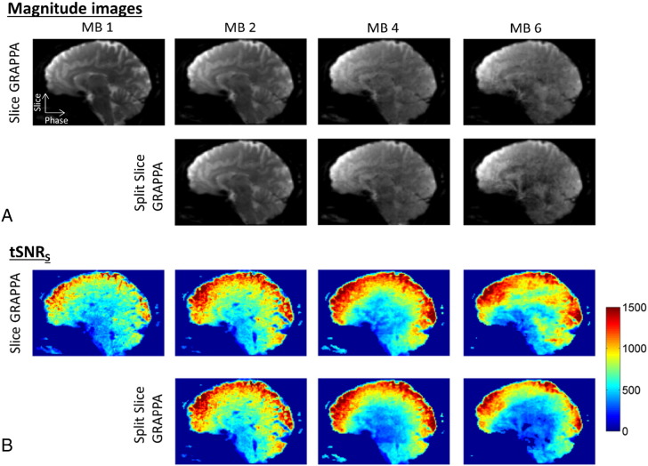
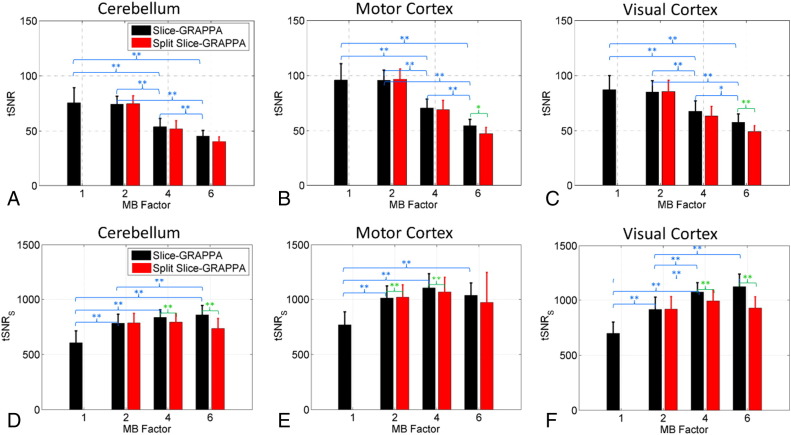
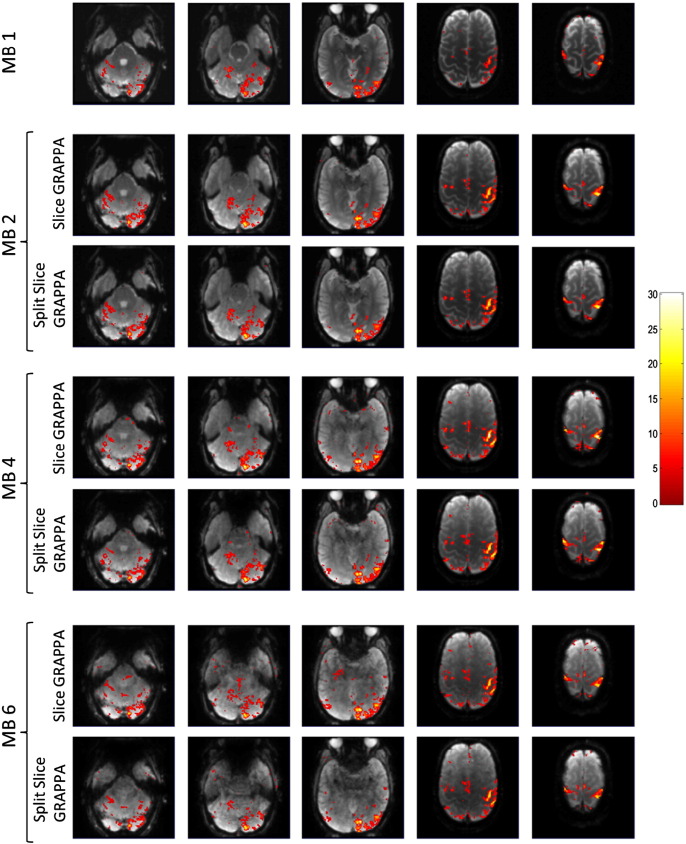
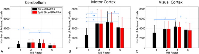
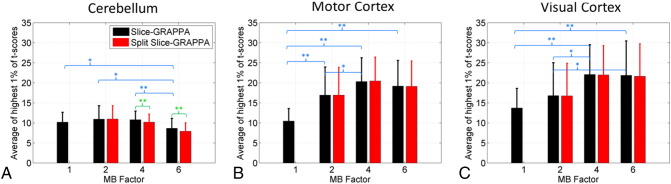
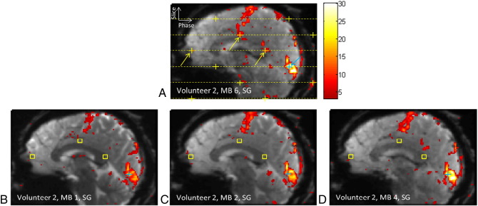
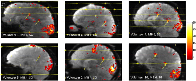
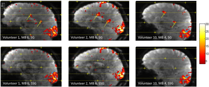
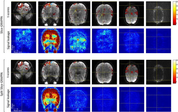
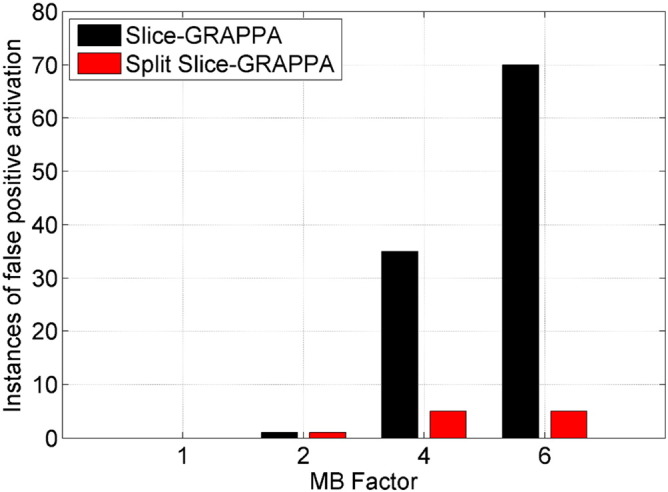
Functional magnetic resonance imaging (fMRI) studies that require high-resolution whole-brain coverage have long scan times that are primarily driven by the large number of thin slices acquired. Two-dimensional multiband echo-planar imaging (EPI) sequences accelerate the data acquisition along the slice direction and therefore represent an attractive approach to such studies by improving the temporal resolution without sacrificing spatial resolution. In this work, a 2D multiband EPI sequence was optimized for 1.5mm isotropic whole-brain acquisitions at 3T with 10 healthy volunteers imaged while performing simultaneous visual and motor tasks. The performance of the sequence was evaluated in terms of BOLD sensitivity and false-positive activation at multiband (MB) factors of 1, 2, 4, and 6, combined with in-plane GRAPPA acceleration of 2× (GRAPPA 2), and the two reconstruction approaches of Slice-GRAPPA and Split Slice-GRAPPA. Sensitivity results demonstrate significant gains in temporal signal-to-noise ratio (tSNR) and t-score statistics for MB 2, 4, and 6 compared to MB 1. The MB factor for optimal sensitivity varied depending on anatomical location and reconstruction method. When using Slice-GRAPPA reconstruction, evidence of false-positive activation due to signal leakage between simultaneously excited slices was seen in one instance, 35 instances, and 70 instances over the ten volunteers for the respective accelerations of MB 2×GRAPPA 2, MB 4×GRAPPA 2, and MB 6×GRAPPA 2. The use of Split Slice-GRAPPA reconstruction suppressed the prevalence of false positives significantly, to 1 instance, 5 instances, and 5 instances for the same respective acceleration factors. Imaging protocols using an acceleration factor of MB 2×GRAPPA 2 can be confidently used for high-resolution whole-brain imaging to improve BOLD sensitivity with very low probability for false-positive activation due to slice leakage. Imaging protocols using higher acceleration factors (MB 3 or MB 4×GRAPPA 2) can likely provide even greater gains in sensitivity but should be carefully optimized to minimize the possibility of false activations.
Keywords: High resolution; Multiband excitation; Simultaneous multislice; Whole brain; fMRI.
Copyright © 2015 The Authors. Published by Elsevier Inc. All rights reserved.
Figures
Similar articles
-
Evaluation of highly accelerated simultaneous multi-slice EPI for fMRI.Neuroimage. 2015 Jan 1;104:452-9. doi: 10.1016/j.neuroimage.2014.10.027. Epub 2014 Oct 18. Neuroimage. 2015. PMID: 25462696 Free PMC article.
-
Accelerated spin-echo functional MRI using multisection excitation by simultaneous spin-echo interleaving (MESSI) with complex-encoded generalized slice dithered enhanced resolution (cgSlider) simultaneous multislice echo-planar imaging.Magn Reson Med. 2020 Jul;84(1):206-220. doi: 10.1002/mrm.28108. Epub 2019 Dec 16. Magn Reson Med. 2020. PMID: 31840295 Free PMC article.
-
Impacts of simultaneous multislice acquisition on sensitivity and specificity in fMRI.Neuroimage. 2018 May 15;172:538-553. doi: 10.1016/j.neuroimage.2018.01.078. Epub 2018 Feb 3. Neuroimage. 2018. PMID: 29408461
-
Rapid brain MRI acquisition techniques at ultra-high fields.NMR Biomed. 2016 Sep;29(9):1198-221. doi: 10.1002/nbm.3478. Epub 2016 Feb 2. NMR Biomed. 2016. PMID: 26835884 Free PMC article. Review.
-
Tradeoffs in pushing the spatial resolution of fMRI for the 7T Human Connectome Project.Neuroimage. 2017 Jul 1;154:23-32. doi: 10.1016/j.neuroimage.2016.11.049. Epub 2016 Nov 25. Neuroimage. 2017. PMID: 27894889 Free PMC article. Review.
Cited by
-
Separating neuronal activity and systemic low-frequency oscillation related BOLD responses at nodes of the default mode network during resting-state fMRI with multiband excitation echo-planar imaging.Front Neurosci. 2022 Sep 21;16:961686. doi: 10.3389/fnins.2022.961686. eCollection 2022. Front Neurosci. 2022. PMID: 36213741 Free PMC article.
-
Task-Dependent Functional and Effective Connectivity during Conceptual Processing.Cereb Cortex. 2021 Jun 10;31(7):3475-3493. doi: 10.1093/cercor/bhab026. Cereb Cortex. 2021. PMID: 33677479 Free PMC article.
-
Simultaneous multi-slice (SMS) acquisition enhances the sensitivity of hemodynamic mapping using gas challenges.NMR Biomed. 2016 Nov;29(11):1511-1518. doi: 10.1002/nbm.3600. Epub 2016 Sep 6. NMR Biomed. 2016. PMID: 27598821 Free PMC article.
-
Neurocognitive Signatures of Naturalistic Reading of Scientific Texts: A Fixation-Related fMRI Study.Sci Rep. 2019 Jul 23;9(1):10678. doi: 10.1038/s41598-019-47176-7. Sci Rep. 2019. PMID: 31337859 Free PMC article.
-
Characterizing functional pathways of the human olfactory system.Elife. 2019 Jul 24;8:e47177. doi: 10.7554/eLife.47177. Elife. 2019. PMID: 31339489 Free PMC article.
References
-
- Ashburner J., Friston K.J. Unified segmentation. NeuroImage. 2005;26:839–851. - PubMed
-
- Bishop J.E., Plewes D.B. TE interleaving: new multisection imaging technique. J. Magn. Reson. Imaging. 1991;1:531–538. - PubMed
-
- Breuer F.A., Blaimer M., Heidemann R.M., Mueller M.F., Griswold M.A., Jakob P.M. Controlled aliasing in parallel imaging results in higher acceleration (CAIPIRINHA) for multi-slice imaging. Magn. Reson. Med. 2005;53:684–691. - PubMed
-
- Crooks L., Arakawa M., Hoenninger J., Watts J., McRee R., Kaufman L., Davis P.L., Margulis A.R., DeGroot J. Nuclear magnetic resonance whole-body imager operating at 3.5 KGauss. Radiology. 1982;143:169–174. - PubMed
Publication types
MeSH terms
Grants and funding
LinkOut - more resources
Full Text Sources
Other Literature Sources
Miscellaneous
