. 2015 Oct:82:574-579.
doi: 10.1016/j.nbd.2015.08.022. Epub 2015 Sep 1.

Neuroprotective effects of urate are mediated by augmenting astrocytic glutathione synthesis and release

Affiliations

Neuroprotective effects of urate are mediated by augmenting astrocytic glutathione synthesis and release

Rachit Bakshi et al. Neurobiol Dis. 2015 Oct.

Abstract

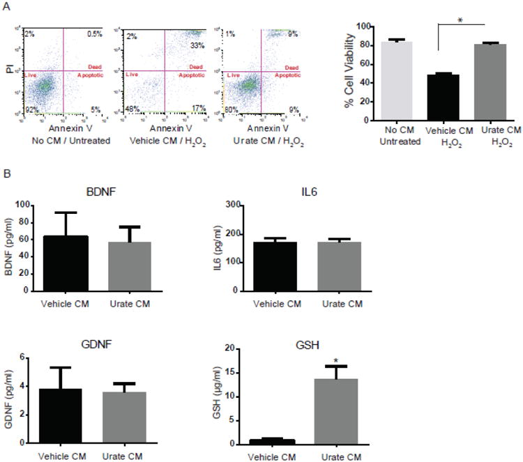
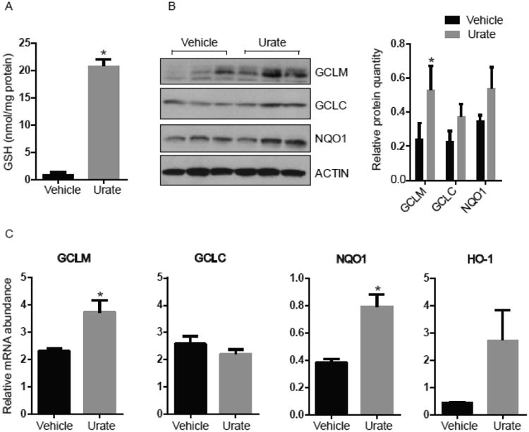
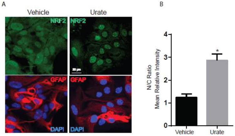
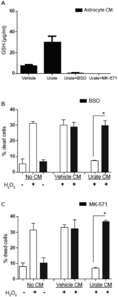
Urate has emerged as a promising target for neuroprotection based on epidemiological observations, preclinical models, and early clinical trial results in multiple neurologic diseases, including Parkinson's disease (PD). This study investigates the astrocytic mechanism of urate's neuroprotective effect. Targeted biochemical screens of conditioned medium from urate- versus vehicle-treated astrocytes identified markedly elevated glutathione (GSH) concentrations as a candidate mediator of urate's astrocyte-dependent neuroprotective effects. Urate treatment also induced the nuclear translocation of the nuclear factor (erythroid-derived 2)-like 2 (Nrf2) protein and transcriptional activation of its key target genes in primary astrocytic cultures. Urate's neuroprotective effect was attenuated when GSH was depleted in the conditioned media either by targeting its synthesis or release by astrocytes. Overall, these results implicate GSH as the extracellular astrocytic factor mediating the protective effect of urate in a cellular model of PD. These results also show that urate can employ a novel indirect neuroprotective mechanism via induction of the Nrf2 signaling pathway, a master regulator of the response to oxidative stress, in astrocytes.

Keywords: Astrocytes; Glutathione; Neurons; Nrf2; Urate.


Figures

Figure 1
Figure 1. Marked elevation in GSH release from astrocytes treated with urate
(A) Conditioned medium (CM) from urate-treated astrocytes protects dopaminergic cells from H2O2-induced cell death. Representative graphs of FACS analysis show cell viability using propidium iodide (PI)/Annexin V staining. Percentages of PI+/AnnexinV+ (dead), PI-/AnnexinV+ (apoptotic) and PI–/AnnexinV– (vital) staining are shown for untreated MES 23.5 cells, or those treated with CM from vehicle- or urate (100 μM)-treated astrocytes a day before and during 200μM H2O2 treatment for 24h. (B) Targeted screening of several neurotrophic factors in the CM. There was no change detected in levels of BDNF, GDNF and IL6 factors in the urate versus vehicle CM. GSH content was significantly increased in CM from urate- (versus vehicle-) treated astrocyte. *denotes p value < 0.001; (n=4 independent experiments).
Figure 2
Figure 2. Urate induces the Nrf2 pathway in astrocytes
(A) GSH levels are elevated in lysates from 100 μM urate- (versus vehicle-) treated astrocytes. (B) Urate induces protein products of Nrf2-targeted genes as shown by western blots of GCLM, GCLC and NQO-1 proteins in urate-treated astrocytes compared to controls. The approximate molecular weight of the indicated protein in kDa is indicated at Left. The graph represents densitometric analysis of the western blots to semi quantify the protein levels. (C) Quantitative PCR analysis of Nrf2 target genes in urate- (versus vehicle-) treated cells. Mean ± SEM are shown (n = 3). * denotes p value < 0.05.
Figure 3
Figure 3. Urate induces nuclear translocation of the Nrf2 protein
(A) Astrocytes were incubated with urate (100 μM) or vehicle control for 8h. Cells were immunostained for astrocyte-specific marker GFAP (red) and nuclei were stained with DAPI (blue). Nrf2 was detected using FITC (green) staining. These representative images show a predominant nuclear distribution of the Nrf2 protein after urate treatment, in contrast to the greater proportion of cytoplasmic expression of Nrf2 in vehicle-treated astrocytes. (B) The graph represents the quantification of the nuclear:cytoplasmic (N/C) ratios of Nrf2 staining intensity using ImageJ software. 20-30 cells from 4 independent experiments were counted. * denotes p value < 0.05.
Figure 4
Figure 4. GSH depletion attenuated the protective effects of urate
(A) GSH content was undetectable in CM from astrocytes that were treated with urate as well as either BSO (0.25 mM) or MK-571 (50 μM). (B,C) MES 23.5 cells were exposed to an oxidative stressor (200 μM H2O2) after pretreatment with CM. The CM was from vechicle- or urate-treated astrocytes, with the latter also treated with or without BSO (0.25 mM) or MK-571 (50 μM) as indicated.. The protective effect of CM from urate-treated astrocytes against H2O2 toxicity (% dead cells) was significantly reduced by astrocyte incubation with BSO (B) or MK-571 (C) (n = 3). * denotes p value < 0.05.

References

    1. Ascherio A, et al. Urate as a predictor of the rate of clinical decline in Parkinson disease. Arch Neurol. 2009;66:1460–8. - PMC - PubMed
    1. Bakshi R, et al. Purines in Parkinson's: Adenosine A2A receptors and urate as targets for neuroprotection. In: Morelli M, editor. The Adenosinergic system - a non-dopaminergic target in Parkinson's disease. Springer; 2015.
    1. Behbahani H, et al. Flow cytometry as a method for studying effects of stressors on primary rat neurons. J Neurosci Res. 2005;82:432–41. - PubMed
    1. Belanger M, Magistretti PJ. The role of astroglia in neuroprotection. Dialogues Clin Neurosci. 2009;11:281–95. - PMC - PubMed
    1. Cai J, et al. Differential regulation of gamma-glutamylcysteine synthetase heavy and light subunit gene expression. Biochem J. 1997;326(Pt 1):167–72. - PMC - PubMed

Publication types

MeSH terms

LinkOut - more resources