. 2015 Dec;100(12):1526-33.
doi: 10.3324/haematol.2015.132183. Epub 2015 Sep 4.

Hemoglobin interaction with GP1bα induces platelet activation and apoptosis: a novel mechanism associated with intravascular hemolysis

Affiliations

Hemoglobin interaction with GP1bα induces platelet activation and apoptosis: a novel mechanism associated with intravascular hemolysis

Rashi Singhal et al. Haematologica. 2015 Dec.

Abstract

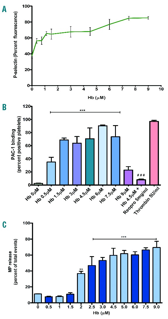
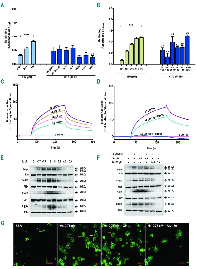
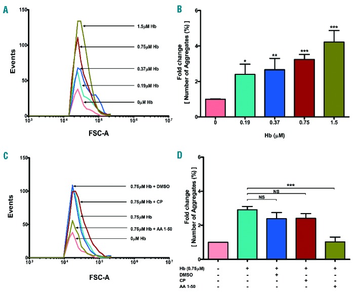
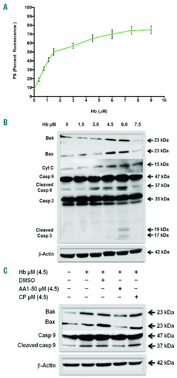
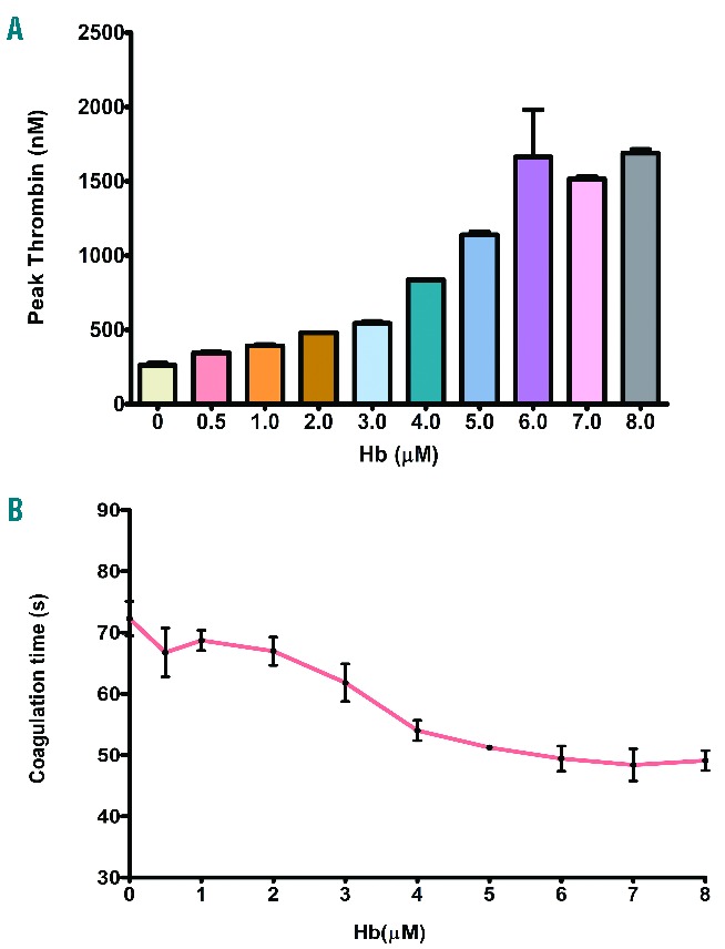
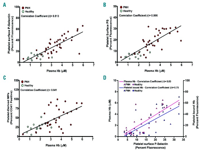
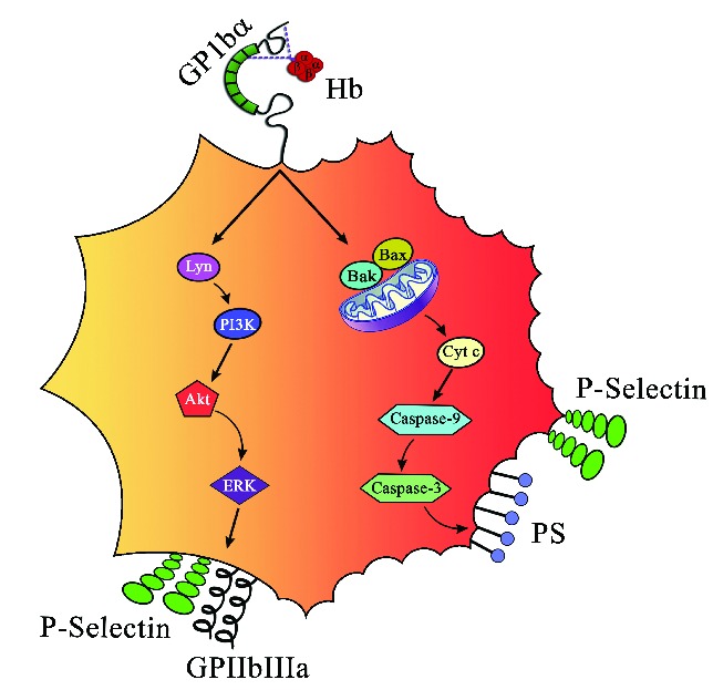
Intravascular hemolysis increases the risk of hypercoagulation and thrombosis in hemolytic disorders. Our study shows a novel mechanism by which extracellular hemoglobin directly affects platelet activation. The binding of Hb to glycoprotein1bα activates platelets. Lower concentrations of Hb (0.37-3 μM) significantly increase the phosphorylation of signaling adapter proteins, such as Lyn, PI3K, AKT, and ERK, and promote platelet aggregation in vitro. Higher concentrations of Hb (3-6 μM) activate the pro-apoptotic proteins Bak, Bax, cytochrome c, caspase-9 and caspase-3, and increase platelet clot formation. Increased plasma Hb activates platelets and promotes their apoptosis, and plays a crucial role in the pathogenesis of aggregation and development of the procoagulant state in hemolytic disorders. Furthermore, we show that in patients with paroxysmal nocturnal hemoglobinuria, a chronic hemolytic disease characterized by recurrent events of intravascular thrombosis and thromboembolism, it is the elevated plasma Hb or platelet surface bound Hb that positively correlates with platelet activation.


Figures

Figure 1.
Figure 1.
(A) Platelet P-selectin expression in the presence of Hb. Washed platelets were incubated with various concentrations of Hb in buffer; labeled with FITC P-selectin antibody and analyzed by flow cytometry. Data presented as mean ± SEM from 3 independent experiments. Hb increased the expression of P-selectin in a concentration-dependent manner (P<0.001). (B) PAC-1 binding to platelet in the presence of Hb. Washed platelets were incubated with various concentrations of Hb. The PAC-1 binding was measured using flow cytometry. The Hb increased the PAC-1 binding in a concentration-dependent manner, ***P<0.0001 (compared to Hb 0 μM) and ###P<0.0001 (Hb 4.5 μM alone). (C) Microparticle generation by platelets treated with Hb. Washed platelets were incubated with various concentrations of Hb. The microparticles (MPs) were analyzed in a flow cytometer. Hb increased the MP generation in a concentration-dependent manner, **P<0.001 and ***P<0.0001 (compared to Hb 0 μM).
Figure 2.
Figure 2.
(A) Hb binding to platelet GP1bα: Effect of antibodies. In ELISA, Hb was incubated with immobilized platelets in the presence of 10 μg/mL of GP1bα or GPIIb-IIIa antibodies. Hb binding was detected using anti-Hb HRP antibody. Data are the mean absorbance ± SD from 3 independent experiments. Hb bound in a concentration-dependent manner, ****P<0.0001. Specifically, AN51, HIP1, and AK2 inhibited the binding with ***P<0.0007, **P<0.004, or ##P<0.0045 compared to Hb 0.15 μM alone. (B) Hb binding to glycocalicin: effect of peptides. In glycocalicin-coated ELISA plate, Hb was incubated. Hb bound to glycocalicin in a concentration dependent manner, ***P<0.0001. When Hb (0.75 μM) was incubated in the presence of equimolar peptides (designed from GP1bα N-terminal), the peptides inhibited Hb binding with **P<0.002, P<0.0017, ##P<0.0045, or NS (non-significant). (C) Hb binding to glycocalicin under flow. Hb was perfused under flow over glycocalicin-coated CM5 chip and binding was measured using BIAcore. Hb bound to glycocalicin with KD = 44 μM. (D) Hb binding to glycocalicin under flow inhibited by AA1-50. Hb (40 μM) was perfused under flow with and without AA1-50 (9 μM). The AA1-50 blocked completely Hb binding to glycocalicin. (E) Hb binding activates platelets. Washed platelets were incubated with various concentrations of Hb. The platelet lysate was immunoblotted for p/non-p-Lyn, PI3K, AKT, or ERK. The Hb (0.37–3.0 μM) increased phosphorylation of the above proteins. Densitometry data are available in Online Supplementary Figure S3. (F) AA1-50 inhibits platelet activation. Washed platelets were incubated with Hb in the presence of control peptide (CP) or AA1-50. The expression of above signaling proteins was measured by immunoblotting. The peptide AA1-50 inhibited the phosphorylation of above proteins dose dependently. Densitometry data are available in Online Supplementary Figure S4. (G) AA1-50 inhibits platelet spreading on immobilized Hb. Washed platelets were incubated over surface coated with Hb (0.75 μM) in the presence of CP or AA1-50. Stained with anti-P selectin FITC. The peptide AA1-50 inhibited the platelet spreading. Arrows indicate the filopodia of spread platelets.
Figure 3.
Figure 3.
Hb mediated platelet aggregation. Washed platelets were incubated with various concentrations of Hb (0–1.5 μM) and stirred in the presence of fibrinogen (500 μg/mL). The aggregates were measured using flow cytometry (method described in Online Supplementary Figure S5), represented as (A) events and (B) percent aggregation (mean ± SEM) from 3 different experiments, *P<0.01, **P<0.001, ***P< 0.002. AA1-50 inhibits aggregation. The above aggregation was performed in the presence of equimolar Hb and peptides (0.75 μM). Data measured as mentioned above, represented as (C) events and (D) percent aggregation. The peptide AA1-50 significantly decreased the Hb-mediated platelet aggregation, ***P< 0.001, NS: non-significant.
Figure 4.
Figure 4.
(A) PS expression in platelets treated with Hb. Washed platelets were incubated with various concentrations of Hb in buffer. Labeled with PE-Cy5 annexin V and analyzed by flow cytometry. The Hb increased PS expression in a concentration-dependent manner, P<0.001. (B) Expression of proapoptotic proteins in platelets in the presence of Hb. Washed platelets were incubated with various concentrations of Hb. The platelet lysate was immunoblotted for Bak, Bax, cytochrome C, caspase-9, and caspase-3. Hb concentrations, 4.5 and 6 Μm, showed increased expression of above proteins including the cleavage upon activation of caspase-9 and caspase-3. The β-Actin is the loading control. Densitometry data are available in Online Supplementary Figure S8. (C) AA1-50 blocks platelet apoptosis. Washed platelets were incubated with Hb in the presence of AA1-50 or CP. The expression of Bak, Bax, and caspase-9 were measured by immunoblotting. The peptide AA1-50 inhibited the expression of above proteins. Densitometry data are available in Online Supplementary Figure S9.
Figure 5.
Figure 5.
(A) Thrombin generation by platelets treated with Hb. Washed platelets treated with Hb have increased thrombin generation dose dependently as measured by the cleavage of fluorogenic substrate by thrombin. The thrombin levels are the mean ± SEM from 3 independent experiments (P<0.001). (B) Coagulation reaction by platelets treated with Hb. Washed platelets treated with Hb were incubated with platelet-poor plasma and coagulation reagents. Data show the mean ± SEM of clot formation time from 3 independent experiments. Hb increased faster coagulation in a concentration-dependent manner (P<0.001).
Figure 6.
Figure 6.
Correlation between plasma Hb/platelet-surface Hb and platelet surface P-selectin, or PS, or platelet-derived MPs. Extracellular Hb concentration was measured from plasma of PNH patients and healthy individuals using sandwich ELISA. Plasma Hb shows positive correlations with (A) P-selectin and (B) PS expression on platelets (freshly isolated from individuals) measured by flow cytometry using FITC-CD62P or PE-Cy5 annexin V antibody. (C) Plasma Hb also correlates positively with platelet-derived microparticles (MPs) in plasma, measured by flow cytometry using CD41-PE antibody. (D) Both platelet surface-bound Hb and plasma extracellular Hb correlates positively with platelet surface P-selectin. Each dot represents individual data. (A–C) Data from 36 PNH patients and 13 healthy controls. (D) Data from another pool of 20 PNH and 10 healthy controls.
Figure 7.
Figure 7.
Schematic representation of platelet activation and apoptosis by Hb. Hb at lower concentrations (0.37–3 μM) induces inside out signaling via Lyn, PI3K, AKT, and ERK by binding to GP1bα and increases surface expression of P-selectin and GPIIb-IIIa. On the other hand, concentrations of Hb (3-6 μM) induce apoptosis and increase expression of Bak and Bax, release cytochrome C, activate caspase-9 and caspase-3, and expose PS on platelets.

Similar articles

Cited by

References

    1. Rother RP, Bell L, Hillmen P, Gladwin MT. The clinical sequelae of intravascular hemolysis and extracellular plasma hemoglobin: a novel mechanism of human disease. JAMA. 2005;293(13):1653–1662. - PubMed
    1. Kristiansen M, Graversen JH, Jacobsen C, et al. Identification of the haemoglobin scavenger receptor. Nature. 2001;409(6817): 198–201. - PubMed
    1. Cappellini MD. Coagulation in the pathophysiology of hemolytic anemias. Hematology Am Soc Hematol Educ Program. 2007:74–78. - PubMed
    1. Barker JE, Wandersee NJ. Thrombosis in heritable hemolytic disorders. Curr Opin Hematol. 1999;6(2):71–75. - PubMed
    1. Regoeczi E, Rubenberg ML, Brain MC. Intravascular haemolysis and disseminated intravascular coagulation. Lancet. 1967;1(7490):601–602. - PubMed

Publication types