. 2015 Sep;27(9):2560-81.
doi: 10.1105/tpc.15.00393. Epub 2015 Sep 4.

ESCRT-III-Associated Protein ALIX Mediates High-Affinity Phosphate Transporter Trafficking to Maintain Phosphate Homeostasis in Arabidopsis

Affiliations

ESCRT-III-Associated Protein ALIX Mediates High-Affinity Phosphate Transporter Trafficking to Maintain Phosphate Homeostasis in Arabidopsis

Ximena Cardona-López et al. Plant Cell. 2015 Sep.

Erratum in

Abstract

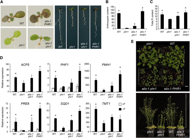
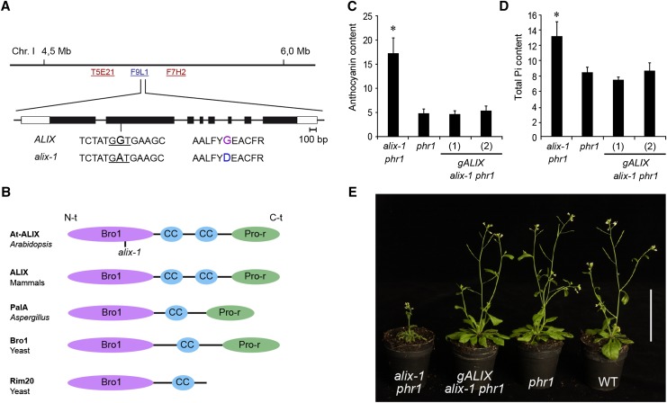
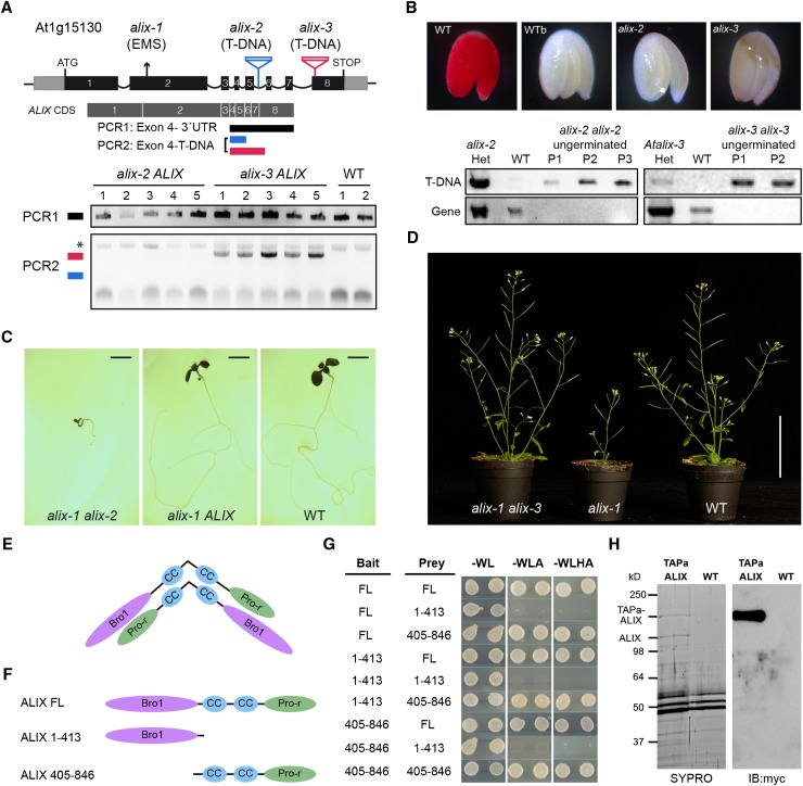
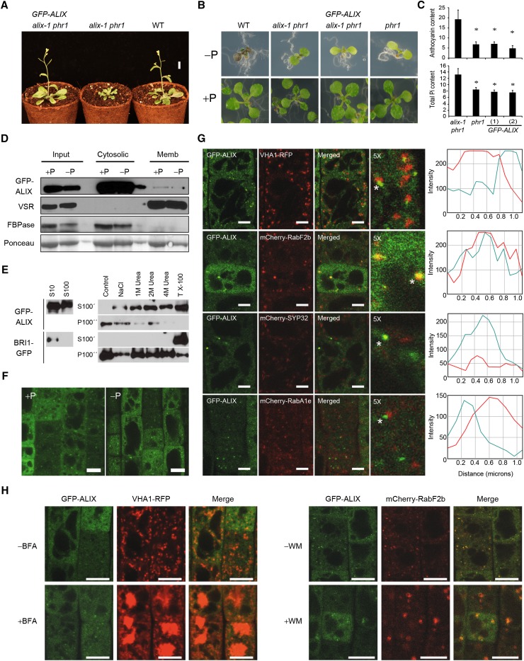
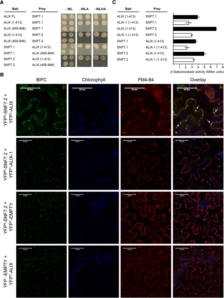
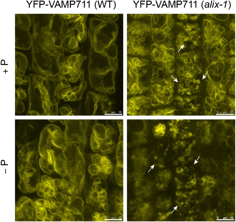
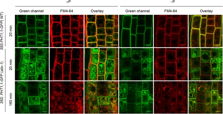
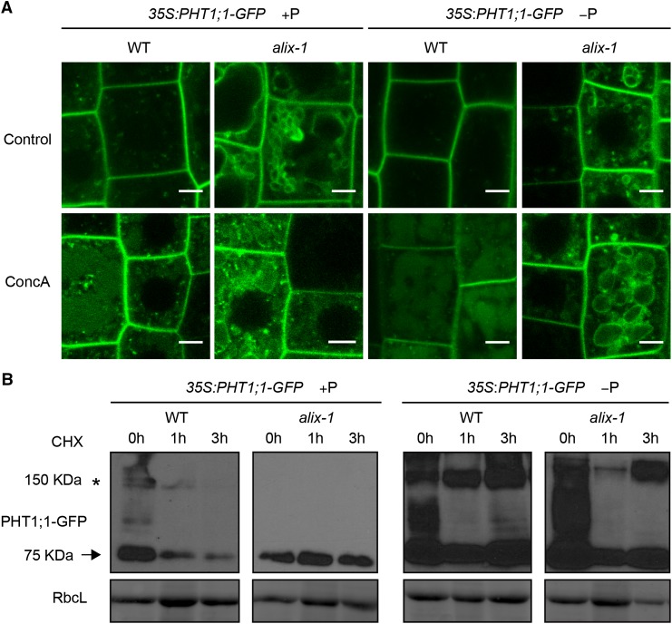
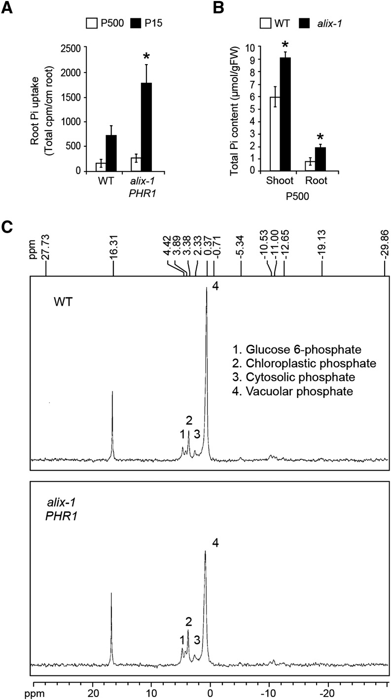
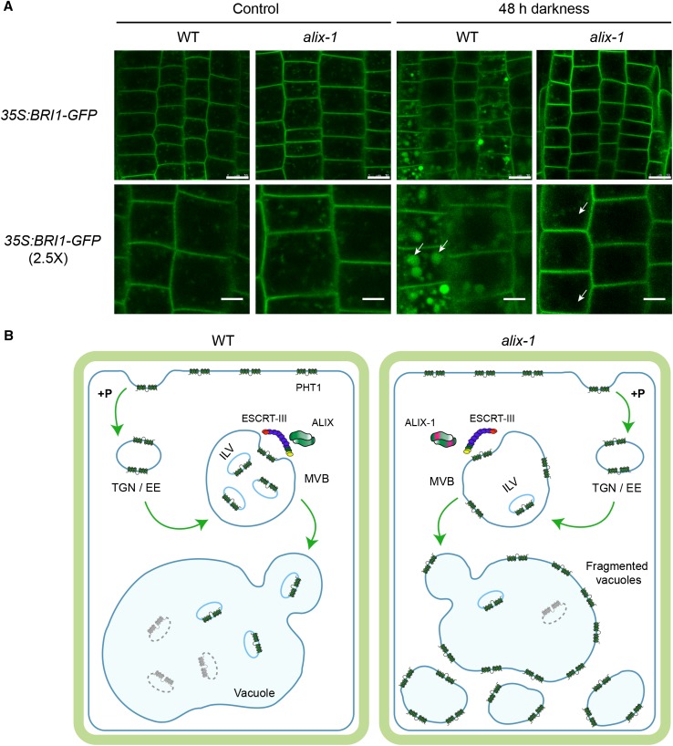
Prior to the release of their cargoes into the vacuolar lumen, sorting endosomes mature into multivesicular bodies (MVBs) through the action of ENDOSOMAL COMPLEX REQUIRED FOR TRANSPORT (ESCRT) protein complexes. MVB-mediated sorting of high-affinity phosphate transporters (PHT1) to the vacuole limits their plasma membrane levels under phosphate-sufficient conditions, a process that allows plants to maintain phosphate homeostasis. Here, we describe ALIX, a cytosolic protein that associates with MVB by interacting with ESCRT-III subunit SNF7 and mediates PHT1;1 trafficking to the vacuole in Arabidopsis thaliana. We show that the partial loss-of-function mutant alix-1 displays reduced vacuolar degradation of PHT1;1. ALIX derivatives containing the alix-1 mutation showed reduced interaction with SNF7, providing a simple molecular explanation for impaired cargo trafficking in alix-1 mutants. In fact, the alix-1 mutation also hampered vacuolar sorting of the brassinosteroid receptor BRI1. We also show that alix-1 displays altered vacuole morphogenesis, implying a new role for ALIX proteins in vacuolar biogenesis, likely acting as part of ESCRT-III complexes. In line with a presumed broad target spectrum, the alix-1 mutation is pleiotropic, leading to reduced plant growth and late flowering, with stronger alix mutations being lethal, indicating that ALIX participates in diverse processes in plants essential for their life.


Figures

Figure 1.
Figure 1.
Molecular and Physiological Characterization of alix-1 Mutants. (A) Photographs of wild-type (WT), phr1, alix-1 phr1, and alix-1 PHR1 plants grown in Pi-deficient medium for 10 d. (B) and (C) Histograms showing anthocyanin and Pi content in wild-type, phr1, alix-1 phr1, and alix-1 PHR1 plants. Seedlings were grown for 12 d in Pi-deficient medium for anthocyanin measurements (Abs 530 nm/g fresh weight; n = 8) and for 10 d in complete medium for Pi content analysis (μM/g fresh weight; n = 6). Error bars indicate standard deviations. (D) RT-qPCR analysis of the expression of representative PSI genes in wild-type, phr1, alix-1 phr1, and alix-1 PHR1 seedlings grown under low Pi (−P; 30 µM Pi) and Pi-sufficient (+P; 500 µM Pi) conditions. ACTIN8 was used as a housekeeping reference gene. Expression levels are relative to Pi-rich-grown wild-type values, which were normalized to 1. Data represent the mean of three biological replicates with sd. (E) Pictures of 28-d-old (up) and 49-d-old (down) wild-type, phr1, alix-1 phr1, and alix-1 PHR1 plants grown in soil. Bars = 1 cm. *P < 0.05 (Student’s t test) with respect to the phr1 mutant in the same experimental conditions.
Figure 2.
Figure 2.
Positional Cloning of the ALIX Gene and the Characteristics of Its Protein. (A) Position of the ALIX locus on chromosome 1 of Arabidopsis (BAC F9L1). The sequence surrounding the alix-1 mutation (G-to-A transition), as well as the resulting amino acid change (Gly260-to-Asp), is shown. The exon structure of ALIX is represented with boxes (light, untranslated; dark, coding region). (B) Diagrams depicting the domain organization of At-ALIX and homolog proteins in mammals (ALIX), A. nidulans (PalA), and yeast (Bro1 and Rim20). ALIX-related proteins comprise a Bro1 domain (Bro1) in the N-terminal region (N-t), followed by one or two coiled-coil (CC) domains and a proline-rich (Pro-r) motif in the C-terminal region (C-t). The position of the alix-1 mutation in the Bro1 domain of At-ALIX protein is indicated. (C) and (D) Complementation of alix-1 mutant defects using a construct containing the ALIX genomic region. Plants corresponding to phr1, alix-1 phr1, and alix-1 phr1 transformed with a construct containing the ALIX genomic region (gALIX alix-1 phr1) were grown for 12 d in Pi-deficient medium for anthocyanin measurements (Abs 530 nm/g fresh weight; n = 8) and for 10 d in complete medium for Pi content analysis (μM/g fresh weight; n = 6). Error bars indicate standard deviations. *P < 0.05 (Student’s t test) with respect to the phr1 mutant in the same experimental conditions. (E) Plants corresponding to the same genotypes as in (C), together with wild-type (WT) plants were grown in soil for 45 d under long-day conditions. Bar = 5 cm.
Figure 3.
Figure 3.
ALIX Forms Dimers and Is Essential for Plant Life. (A) A diagram of At-ALIX genomic region showing the position of the EMS-induced G-to-A mutation in alix-1 plants and that of T-DNA insertions in alix-2 and alix-3 mutants. In lower panels, PCR analysis was used to detect potential truncated ALIX transcripts in the cDNA of alix-2 and alix-3 heterozygous mutants (five lines per mutant). An asterisk indicates a nonspecific band amplified in all samples. (B) Genotyping and tetrazolium staining shows that embryos from ungerminated seeds of the alix-2 and alix-3 progenies correspond to homozygous mutants that are not viable. Embryos from imbibed (WT) and boiled wild-type (WTb) seeds were used as a control. PCR analysis of wild-type and alix mutant embryos was performed as described in Methods to detect T-DNA insertions in the ALIX gene. P represents pools of 15 embryos. (C) Photographs of 10-d-old nonviable transheterozygous alix-1 alix-2 and viable heterozygous alix-1 ALIX and wild-type seedlings. Bars = 0.2 cm. (D) Photographs of trans-heterozygous alix-1 alix-3, homozygous alix-1, and wild-type seedlings grown in soil. Bar = 5 cm. (E) Model for hypothetical ALIX dimerization following an antiparallel disposition. (F) and (G) Yeast two-hybrid assays using full-length (FL) and truncated versions (comprising the Bro1 domain, amino acids 1 to 413; or the coiled coils plus the Pro-rich region, amino acids 405 to 846) of ALIX. Transformed yeast cells were grown in SD-WL medium as a transformation control and in SD-WLA and SD-WLHA media for interaction assays. (H) Copurification of TAPa-ALIX and endogenous ALIX proteins. TAPa-purified proteins were separated in a 10% SDS-PAGE gel and subjected to immunoblot analysis using anti-myc. SYPRO staining was used to visualize differentially purified protein bands prior to their mass spectrometry analysis. Wild-type protein extract was used as a TAPa negative control.
Figure 4.
Figure 4.
Functional GFP-ALIX Fusion Protein Localizes in Both Cytosolic and Microsomal Fractions. (A) Plants corresponding to the wild type (WT), alix-1 phr1, and alix-1 phr1 transformed with a construct containing the ALIX genomic region fused to GFP driven by the ALIX promoter (GFP-ALIX) were grown in soil for 3 weeks before photographs were taken. Bar = 1 cm. (B) Plants as in (A) together with phr1 were grown in Pi-deficient and Pi-rich media for 12 d before photographs were taken. (C) Histograms showing anthocyanin and Pi content in phr1, alix-1 phr1, and GFP-ALIX (two independent transgenic lines) plants. Error bars indicate standard deviations. *P < 0.05 (Student’s t test) with respect to the phr1 mutant in the same experimental conditions. (D) Isolation of microsomes from postnuclear fractions (Input) of 10-d-old GFP-ALIX seedlings grown under Pi-rich (+P) and -deficient (−P) conditions. GFP-ALIX was found in the soluble fraction (Cytosolic), which corresponds to cytosol, as well as associated to microsomes (Memb). (E) Membrane association of GFP-ALIX can be disrupted with chaotropic agents and detergents. Supernatant (S10) samples were ultracentrifuged to give soluble fractions (S100) and pellets. Pellets were resuspended in homogenization buffer without additives (control); with 1 M NaCl (NaCl); with 1, 2, or 4 M urea; or with 1% Triton X-100 (TX-100) and ultracentrifuged again, giving wash fractions (S100’) and pellets. This procedure was repeated to give washed pellets (P100’’). Same procedures were followed with BRI1-GFP as a detergent-solubilized control. (F) Confocal images of root epidermal cells from 5-d-old GFP-ALIX seedlings grown under Pi-deficient (−P) and -rich (+P) conditions. Bars = 10 μm. (G) Confocal images of root epidermal cells of 5-d-old seedlings expressing GFP-ALIX and different cell compartment markers; VHA1-RFP (early endosome), mCherry-RabF2b (MVB), mCherry-SYP32 (Golgi), mCherry-RabA1e (recycling endosome). The 5× enlarged images of merged color channels are shown. ImageJ quantification of green (turquoise lines) and red (red lines) signal intensities for spots indicated by asterisks. Bars = 5 μm. (H) BFA (left panels) and WM (right panels) treatments of 5-d-old GFP-ALIX seedlings expressing VHA1-RFP (for BFA) or mCherry-Rabf2b (for WM). Bars = 10 μm.
Figure 5.
Figure 5.
ALIX Interacts with ESCRT-III Complex Component VPS32/SNF7 through Its Bro1 Domain. (A) Yeast two-hybrid assays showing interaction between the Bro1 domain of At-ALIX and SNF7.1 and SNF7.2. ALIX full-length (FL) and truncated versions (comprising the Bro1 domain, amino acids 1 to 413; or the coiled coils plus the Pro-rich region, amino acids 405 to 846) were used. Transformed yeast cells were grown in SD-WL medium as a transformation control and in SD-WLA and SD-WLHA media for interaction assays. (B) BiFC assays show that ALIX, but not a version containing the alix-1 mutation, interacts with SNF7.2 in vivo. FM-4-64 (5 μM) was injected in Nicotiana benthamiana leaf epidermal cells expressing different construct combinations as indicated. Leaves were observed by confocal imaging after 60 min. Reconstitution of YFP fluorescence indicates that the corresponding ALIX and SNF7 constructs directly interact. White arrows show YFP fluorescence colocalization with FM4-64 signal (red channel). Plastid autofluorescence due to chlorophyll is shown in the blue channel. Bars = 50 μm. (C) alix-1 mutation reduces the ability of the Bro1 domain to interact with SNF7 proteins. β-Galactosidase assays were performed on yeast cotransfected with plasmids expressing indicated recombinant proteins. Error bars indicate standard deviations. n = 6.
Figure 6.
Figure 6.
alix-1 Mutants Are Defective in Vacuolar Size and Morphology. Confocal images of root cells from 5-d-old wild-type (WT) and alix-1 mutants overexpressing the tonoplast marker YFP-VAMP711 (Geldner et al., 2009). Increased number of vacuoles of smaller size (shown by arrows) than those in controls can be observed in alix-1 mutants independently of Pi status. Bars = 10 μm.
Figure 7.
Figure 7.
PHT1;1-GFP Is Mislocalized in alix-1 Mutants. Confocal images of root epidermal cells from 5-d-old wild-type (WT) and alix-1 seedlings overexpressing a PHT1;1-GFP fusion grown in +P and −P conditions. Seedlings were treated with 2 μM FM4-64 for 5 min, washed, and visualized after 20 and 180 min. The green and red channels correspond to the PHT1;1-GFP and membrane-associated FM4-64 fluorescence, respectively. Overlay of both channels in images after 180 min shows PHT1;1-GFP localization in alix-1 tonoplasts. Bars = 10 μm.
Figure 8.
Figure 8.
alix-1 Mutation Alters PHT1;1-GFP Degradation. (A) Confocal images of root cells from 5-d-old 35S:PHT1;1-GFP seedlings in wild-type (WT) and alix-1 mutant backgrounds treated with 1 μΜ ConcA for 6 h. Bars = 5 μm. (B) Immunoblots showing PHT1;1-GFP (arrow) degradation over time in 10-d-old 35S:PHT1;1-GFP seedlings in wild-type and alix-1 mutant backgrounds grown in +P or −P conditions, incubated or not during 1 and 3 h with 50 μM cycloheximide. Anti-GFP was used to detect PHT1;1-GFP. Ponceau staining of the large subunit of Rubisco (RbcL) was used as loading control. An asterisk indicates the position of PHT1;1-GFP aggregates as previously reported by Bayle et al. (2011).
Figure 9.
Figure 9.
Pi Uptake and Vacuolar Storage Are Altered in alix-1 Mutants. (A) Phosphate absorption capacity of alix-1 mutants compared with wild-type plants. 33PO4 uptake was measured in plants grown for 12 d in low Pi (15 μM) or Pi-sufficient (500 μM) medium. Data represent the means and standard deviations of results obtained for 15 plants. (B) Pi content analysis in shoots and roots of alix-1 mutants and wild-type plants grown for 12 d in Pi-sufficient (500 μM) medium. Error bars indicate standard deviations. n = 10. *P < 0.05 (Student’s t test) with respect to the wild type in the same experimental conditions. (C) In vivo 31P-NMR spectra from roots of alix-1 and wild-type seedlings grown in Pi-sufficient (500 μM) medium. Peaks from left to right are assigned to glucose-6-phosphate (1), chloroplastic Pi (2), (3) cytoplasmic Pi, and (4) vacuolar Pi.
Figure 10.
Figure 10.
Vacuolar Degradation of BRI1 Is Defective in alix-1 Mutants. (A) Root epidermal cells from 5-d-old seedlings overexpressing BRI1-GFP in wild-type (WT) and alix-1 backgrounds grown in Pi-rich conditions were kept in the dark or in light for 48 h before they were observed by confocal imaging. White arrows point to vacuoles. Bars = 10 μm except in 2.5× zoom images, where bars = 5 μm. (B) Proposed model for defects in PHT1 localization in alix-1 mutants. Under Pi-rich conditions (+P), Pi transporters (PHT1) are readily endocytosed at the plasma membrane and transported to the TGN/EE. TGN/EE vesicles mature to MVBs, where cargo proteins are packaged into ILVs by the action of ESCRT complexes and associated proteins (i.e., ALIX). Finally, MVBs fuse with vacuoles and release ILVs to the vacuole lumen where they are degraded together with their cargoes. In alix-1 mutants, ALIX-1 association with ESCRT-III complexes is altered, and PHT1;1 proteins are not correctly internalized into ILVs, being retained in the tonoplast upon MVB fusion with vacuoles. This effect leads to reduced PHT1 degradation. Defects in cargo trafficking in alix-1 also provoke vacuolar fragmentation, which alters Pi storage and subcellular distribution, and, therefore, Pi homeostasis.

Similar articles

Cited by

References

    1. Ames B.N. (1966). Assay of inorganic phosphate, total phosphate and phosphatases. Methods Enzymol. 8: 115–118.
    1. Aubert S., Alban C., Bligny R., Douce R. (1996). Induction of beta-methylcrotonyl-coenzyme A carboxylase in higher plant cells during carbohydrate starvation: evidence for a role of MCCase in leucine catabolism. FEBS Lett. 383: 175–180. - PubMed
    1. Ayadi A., David P., Arrighi J.F., Chiarenza S., Thibaud M.C., Nussaume L., Marin E. (2015). Reducing the genetic redundancy of Arabidopsis PHOSPHATE TRANSPORTER1 transporters to study phosphate uptake and signaling. Plant Physiol. 167: 1511–1526. - PMC - PubMed
    1. Babst M. (2005). A protein’s final ESCRT. Traffic 6: 2–9. - PubMed
    1. Babst M., Katzmann D.J., Estepa-Sabal E.J., Meerloo T., Emr S.D. (2002a). Escrt-III: an endosome-associated heterooligomeric protein complex required for mvb sorting. Dev. Cell 3: 271–282. - PubMed

Publication types

MeSH terms