. 2015 Oct 16;290(42):25595-608.
doi: 10.1074/jbc.M115.661413. Epub 2015 Sep 4.

Pro-oncogenic Roles of HLXB9 Protein in Insulinoma Cells through Interaction with Nono Protein and Down-regulation of the c-Met Inhibitor Cblb (Casitas B-lineage Lymphoma b)

Affiliations

Pro-oncogenic Roles of HLXB9 Protein in Insulinoma Cells through Interaction with Nono Protein and Down-regulation of the c-Met Inhibitor Cblb (Casitas B-lineage Lymphoma b)

Shruti S Desai et al. J Biol Chem. .

Abstract

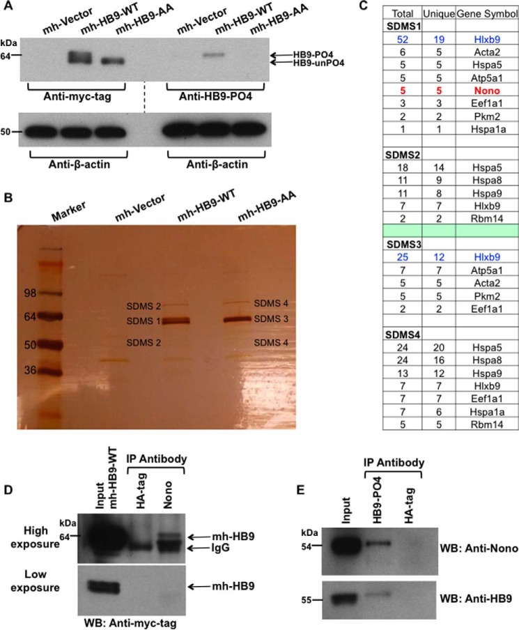
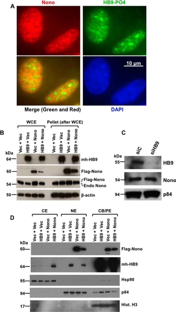
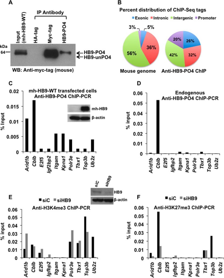
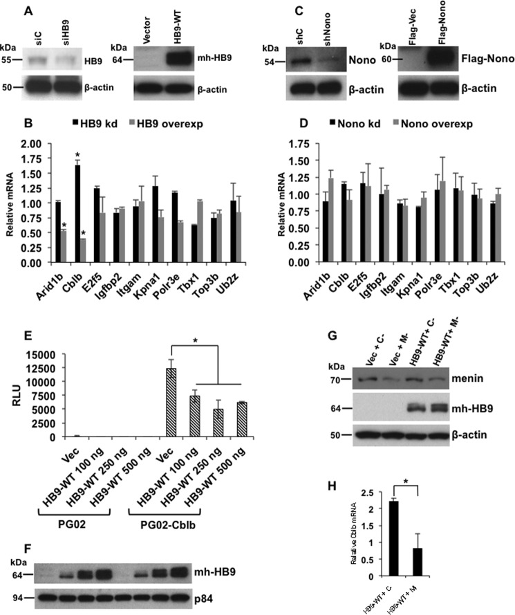
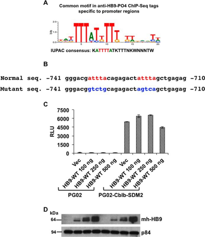
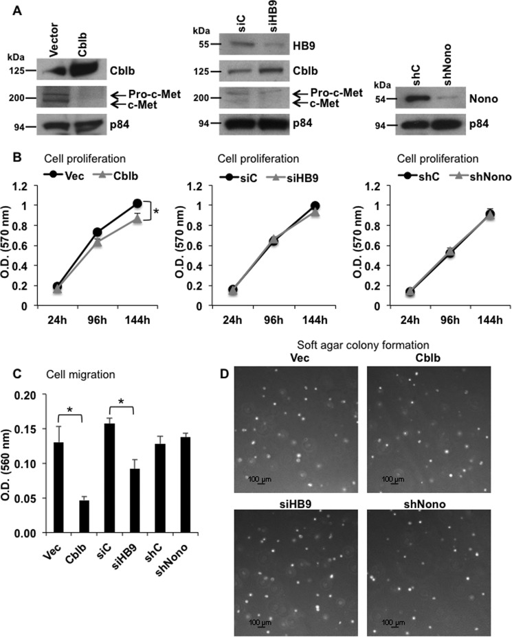
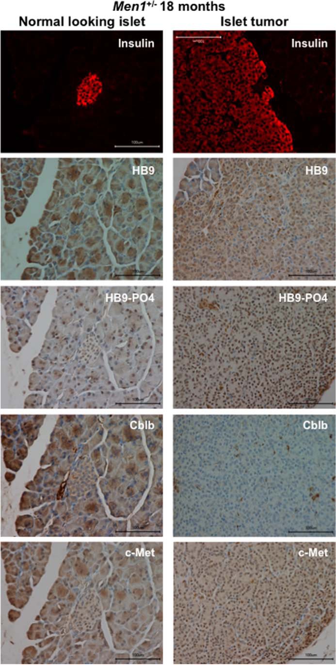
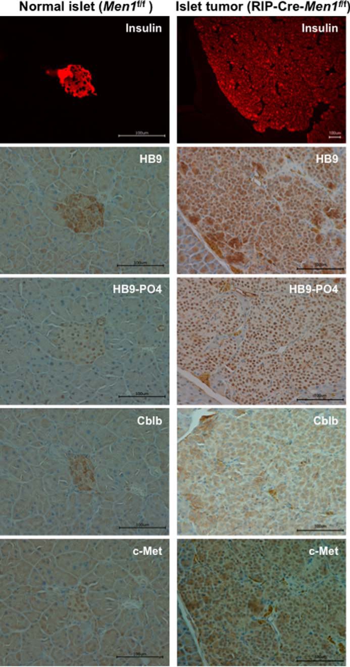
Pancreatic islet β-cells that lack the MEN1-encoded protein menin develop into tumors. Such tumors express the phosphorylated isoform of the β-cell differentiation transcription factor HLXB9. It is not known how phospho-HLXB9 acts as an oncogenic factor in insulin-secreting β-cell tumors (insulinomas). In this study we investigated the binding partners and target genes of phospho-HLXB9 in mouse insulinoma MIN6 β-cells. Co-immunoprecipitation coupled with mass spectrometry showed a significant association of phospho-HLXB9 with the survival factor p54nrb/Nono (54-kDa nuclear RNA-binding protein, non-POU-domain-containing octamer). Endogenous phospho-HLXB9 co-localized with endogenous Nono in the nucleus. Overexpression of HLXB9 decreased the level of overexpressed Nono but not endogenous Nono. Anti-phospho-HLXB9 chromatin immunoprecipitation followed by deep sequencing (ChIP-Seq) identified the c-Met inhibitor, Cblb, as a direct phospho-HLXB9 target gene. Phospho-HLXB9 occupied the promoter of Cblb and reduced the expression of Cblb mRNA. Cblb overexpression or HLXB9 knockdown decreased c-Met protein and reduced cell migration. Also, increased phospho-HLXB9 coincided with reduced Cblb and increased c-Met in insulinomas of two mouse models of menin loss. These data provide mechanistic insights into the role of phospho-HLXB9 as a pro-oncogenic factor by interacting with a survival factor and by promoting the oncogenic c-Met pathway. These mechanisms have therapeutic implications for reducing β-cell proliferation in insulinomas by inhibiting phospho-HLXB9 or its interaction with Nono and modulating the expression of its direct (Cblb) or indirect (c-Met) targets. Our data also implicate the use of pro-oncogenic activities of phospho-HLXB9 in β-cell expansion strategies to alleviate β-cell loss in diabetes.

Keywords: PNET; c-Met; gene regulation; menin (MEN1); neuroendocrine; p54nrb/Nono; pancreatic islet; pathogenesis; phospho-HLXB9; tumor.


Figures

FIGURE 1.
FIGURE 1.
Identification of Nono as a phospho-HLXB9 interacting protein. A, overexpression of HLXB9 shows both its phosphorylated and unphosphorylated isoform. WCE were prepared from MIN6-4N cells transfected with myc-His-tag empty vector (mh-Vector) or plasmids expressing myc-his-tagged HLXB9 (mh-HB9-WT) or the phospho-dead mutant of HLXB9 with alanine substitution at Ser-78 and Ser-80 (mh-HB9-AA). WCE were run on the same gel to generate two Western blots to probe with anti-myc-tag or anti-HB9-PO4 (phospho-HLXB9 antibody). To analyze the bands, the blots were placed side-by-side (indicated by the dotted line). The top band of the doublet in the lane marked mh-HB9-WT corresponds to phospho-HLXB9 because it is not detected with the myc-tag antibody in mh-HB9-AA, and it is detected specifically with anti-HB9-PO4. The bottom band of the doublet corresponds to the unphosphorylated isoform of HLXB9 (HB9-unPO4) because it is not detected with anti-HB9-PO4. β-Actin was used as the loading control. B, large scale co-IP shows the two isoforms of HLXB9 and co-immunoprecipitating proteins. Silver-stained gel of proteins separated on SDS-PAGE after large scale co-IP with a myc-tag antibody using WCE prepared in A. As also seen by Western blot analysis in A, the bands marked SDMS1 and SDMS3 show the doublet in mh-HB9-WT Co-IP (PO4-HB9 and unPO4-HB9) and a single band in mh-HB9-AA corresponding to unPO4-HB9. Bands marked SDMS1, SDMS2, SDMS3, and SDMS4 (that were absent in the mh-Vector lane) were excised from the gel and subjected to mass spectrometry analysis. C, Nono uniquely co-immunoprecipitates with phospho-HLXB9. The number of peptides (total and unique) in the bands excised from the gel shown in B and their corresponding proteins is shown. Several proteins were present in both the mh-HB9-WT and mh-HB9-AA immunoprecipitates. The protein Nono emerged as a phospho-HLXB9-specific partner found only in the co-IP of mh-HB9-WT and not in the co-IP of the phospho-dead mutant of HLXB9 (mh-HB9-AA). D, overexpressed HLXB9 can Co-IP with endogenous Nono. Western blot (WB) probed with anti-myc-tag showing specific co-IP of HLXB9 with Nono using WCE of MIN6-4N cells transfected with mh-HB9-WT. Anti-HA-tag was used as a negative control. Higher exposure (top panel) and lower exposure (bottom panel) of the blot are shown to clearly visualize the input HLXB9 bands. IP, immunoprecipitation. E, endogenous phospho-HLXB9 can co-immunoprecipitate with endogenous Nono. Western blots probed with anti-Nono and anti-HB9 show specific co-IP of endogenous Nono with endogenous phospho-HLXB9 using WCE of MIN6-4N cells. An anti-HA-tag was used as a negative control.
FIGURE 2.
FIGURE 2.
HLXB9 co-localizes with Nono in the nucleus, and co-overexpression of HLXB9 and Nono decreases the overexpression of Nono with translocation of HLXB9 into the cytoplasm. A, endogenous phospho-HLXB9 co-localizes with endogenous Nono in the nucleus. IF images of MIN6-4N cells show endogenous Nono (red) and phospho-HLXB9 (green). DAPI was used to detect the nuclei (blue). A merged image of the red and green IF shows co-localization of Nono and phospho-HLXB9 in subnuclear spots and some regions with phospho-HLXB9 (green) that did not co-localize with Nono. B, overexpression of HLXB9 decreases the level of overexpressed Nono protein. Western blots are shown of WCE and the pellet leftover after WCE preparation from MIN6-4N cells expressing mh-HB9-WT, FLAG-Nono, or both together. Empty vector DNA (Vec) was used to maintain the same amount of DNA in the transfections. The expression of transfected HLXB9 was detected with the anti-myc-tag; Nono was detected with the anti-FLAG-tag and with anti-Nono to detect both endogenous and transfected FLAG-tagged Nono. β-Actin was used as the loading control. Endogenous Nono levels were not affected by HLXB9 overexpression. However, the level of transfected FLAG-Nono was reduced upon HLXB9 overexpression. A similar pattern of bands was seen in the pellet leftover after WCE preparation, indicating that the reduced level of Nono upon HLXB9 overexpression was not due to differential cell lysis in the WCE preparation. C, HLXB9 did not reduce the expression of endogenous Nono protein. Western blot analysis to detect endogenous HLXB9 and Nono using WCE prepared from MIN6-4N cells transfected with control siRNA (siC) or HLXB9 siRNA (siHB9) is shown. p84 was used as the loading control. HLXB9 was significantly knocked down, but that did not affect the level of endogenous Nono. D, co-overexpression of HLXB9 and Nono reduces the level of Nono protein in the nucleus with translocation of HLXB9 to the cytoplasm. Shown is Western blot analysis of subcellular fractionation of CE, NE, and CB/PE from MIN6-4N cells expressing mh-HB9-WT, FLAG-Nono, or both together. Empty vector DNA (Vec) was used to maintain the same amount of DNA in the transfections. The expression of transfected HLXB9 was detected with the anti-myc-tag; Nono was detected with the anti-FLAG-tag; detection of marker proteins (Hsp90 for CE, p84 for NE, and histone H3 for CB/PE) showed minimal cross-contamination of the fractions and also served as loading controls for each fraction. Overexpressed Nono was found in the NE and in the CB/PE, but its level in NE was reduced by HLXB9 overexpression. Overexpressed HLXB9 was mostly located in the CB/PE and with a significant amount in the nucleus, but it was also detected in the CE by Nono overexpression.
FIGURE 3.
FIGURE 3.
Identification of Cblb as a phospho-HLXB9 target gene. A, the anti-HB9-PO4 antibody specifically recognizes the phosphorylated isoform of HLXB9. WCE and chromatin were prepared from MIN6-4N cells transfected with a plasmid expressing myc-his-tagged HLXB9 (mh-HB9-WT). WCE was used IP with rabbit antibodies anti-myc-tag or anti-HB9-PO4 and detected by Western blot (WB) with mouse anti-myc-tag. Rabbit anti-HA-tag was used as the negative control. The input WCE and anti-myc-tag IP display a doublet corresponding to phospho-HLXB9 and unphosphorylated HLXB9. Anti-HB9-PO4 could specifically immunoprecipitate phospho-HLXB9 corresponding to the top band of the doublet. B, significant enrichment of promoter regions among the anti-HB9-PO4 ChIP-Seq tags. Chromatin prepared from MIN6-4N cells transfected in A was used for ChIP with anti-HB9-PO4. DNA obtained before and after ChIP was used for preparing libraries followed by deep sequencing (ChIP-Seq) and mapping of the anti-HB9-PO4-specific ChIP-Seq tags to the mouse genome. The pie chart shows the percent distribution of tags in the mouse genome (a typical input library, Genomatix) and in the anti-HB9-PO4 ChIP-Seq at the indicated regions; 20% of the anti-HB9-PO4 ChIP-Seq tags were located near promoter regions and selected for further analysis. C, phospho-HLXB9 occupancy is highest at the Arid1b and Cblb gene in cells overexpressing HLXB9. ChIP-quantitative PCR assay for validating the 10 phospho-HLXB9 targets is shown as the percent of input chromatin DNA PCR for each primer pair. Chromatin prepared from MIN6-4N cells expressing mh-HB9-WT was used for anti-HB9-PO4 ChIP. Also shown is a Western blot confirming overexpression of HLXB9 (myc-tag antibody) and β-actin as the loading control. D, endogenous phospho-HLXB9 occupancy is highest at the Cblb gene. ChIP-quantitative PCR assay of the 10 phospho-HLXB9 targets is shown as percent of input chromatin DNA PCR for each primer pair. Chromatin prepared from MIN6-4N cells was used for endogenous anti-HB9-PO4 ChIP. Endogenous phospho-HLXB9 occupancy was only detected at Cblb. E and F, H3K4me3 at Cblb unaffected but reduced H3K27me3 upon HLXB9 knockdown. ChIP-quantitative PCR assay of the 10 phospho-HLXB9 targets is shown as the percent of input chromatin DNA PCR for each primer pair. Chromatin prepared from MIN6-4N cells transfected with control siRNA (siC) or HLXB9 siRNA (siHB9) was used for endogenous anti-H3K4me3 ChIP (E) or H3K27me3 ChIP (F). Also shown is a Western blot confirming knockdown of HLXB9 (HLXB9 antibody) and β-actin as the loading control. In siC versus siHB9, reciprocal H3K4me3 or H3K27me3 at only Cblb was HLXB9 binding-dependent because endogenous phospho-HLXB9 was only found to occupy Cblb (D).
FIGURE 4.
FIGURE 4.
HLXB9 suppresses the expression of Cblb mRNA by suppressing Cblb promoter activity. A and B, among targets identified by anti-HB9-PO4 ChIP-Seq, the expression of only Arid1b and Cblb was affected by HLXB9. Quantitative RT-PCR is shown of the indicated genes using RNA prepared from MIN6-4N cells transfected with control siRNA (siC) or HLXB9 siRNA (siHB9), empty vector, or mh-HB9-WT. A, Western blot confirming knockdown or overexpression of HLXB9 with anti-HLXB9 or anti-myc-tag, respectively, and β-actin as the loading control. B, HLXB9 knockdown or overexpression regulated the relative mRNA level of only two genes (Arid1b and Cblb). Both were reduced upon HLXB9 overexpression, but only Cblb was increased upon HLXB9 knockdown. Error bar = mean and S.D. from three experiments. * = p < 0.05. C and D, Nono did not regulate the expression of HLXB9 target genes. Shown is quantitative RT-PCR of the indicated genes using RNA prepared from MIN6-4N cells transfected with control shRNA (shC) or Nono shRNA (shNono), FLAG-vector, or FLAG-Nono. A, Western blot confirming knockdown or overexpression of Nono with anti-Nono or anti-FLAG-tag, respectively, and β-actin as the loading control. B, Nono knockdown or overexpression did not regulate the relative mRNA level of any gene. Error bar = mean and S.D. from three experiments. E and F, Cblb promoter activity is suppressed by HLXB9. The promoter region of Cblb (−1078 to +219) located near the sequence identified by anti-HB9-PO4 ChIP-Seq was cloned in the promoter-less luciferase reporter vector PG02 (GeneCopoeia) and analyzed for promoter activity in MIN6-4N cells. RLU for each of the transfections are shown. Compared with the empty vector PG02, the PG02-Cblb plasmid showed significantly high RLU, and co-expression of increasing amounts of HLXB9 suppressed the Cblb promoter activity. Error bar = mean and S.D. from three experiments. * = p < 0.05. A representative Western blot shows the expression of HLXB9 (with anti-myc-tag) in the MIN6-4N cells analyzed for luciferase activity. p84 was used as the loading control. G and H, Cblb expression is down-regulated by the phosphorylated isoform of HLXB9. WCE and RNA were prepared from MIN6-4N cells transfected with control-shRNA (C−) or Men1-shRNA (M−) together with empty vector or mh-HB9-WT. G, Western blot shows the extent of menin knockdown (anti-menin blot) and transfected HLXB9 and increased phospho-HLXB9 in M− (anti-myc-tag blot, top band of the doublet band). H, quantitative RT-PCR shows significantly reduced Cblb mRNA upon menin knockdown in mh-HB9-WT-transfected cells. Error bar = mean and S.D. of a representative experiment performed in triplicate. * = p < 0.05.
FIGURE 5.
FIGURE 5.
HLXB9 binding motif in the Cblb promoter. A, consensus motif in anti-HB9-PO4 ChIP-Seq tags located at promoter regions. De novo motif analysis from anti-HB9-PO4 ChIP-Seq tag sequences located at promoter regions was performed using Genomatix software. A core sequence ATTTTA was identified that resembles homeodomain-binding consensus (35). B, sequence of the HLXB9 binding motifs in the Cblb promoter. The top line shows the putative HLXB9 binding motifs (red) in the DNA sequence of the Cblb promoter (−741 to −710 region from the transcriptional start site is shown). The bottom line shows nucleotide substitutions (blue) to mutate the motifs by site-directed mutagenesis in the Cblb-promoter construct used in C. C and D, HLXB9 did not suppress the activity of the Cblb promoter containing mutations at the HLXB9 binding motifs. The putative HLXB9 binding motifs shown in B were mutated by site-directed mutagenesis of the PG02-Cblb promoter construct and analyzed for promoter activity in MIN6-4N cells. RLU for each of the transfections are shown. Compared with the empty vector PG02, the PG02-Cblb-SDM2 plasmid showed significantly high RLU and co-expression of increasing amounts of HLXB9 did not suppress the Cblb promoter activity. Error bar = Mean and S.D. from 3 experiments, * = p < 0.05. A representative Western blot shows expression of HLXB9 (with anti-myc-tag) in the MIN6-4N cells analyzed for luciferase activity. p84 was used as the loading control.
FIGURE 6.
FIGURE 6.
Cblb overexpression or HLXB9 knockdown inactivates the oncogenic c-Met pathway. A, overexpression of Cblb or knockdown of HLXB9 decreases c-Met levels. Western blot analysis of the indicated proteins using WCE prepared from MIN6-4N cells transfected (by nucleofection) with empty vector or Cblb expression plasmid, control siRNA (siC), or HLXB9 siRNA (siHB9) and control shRNA (shC) or Nono shRNA (shNono). Cblb overexpression reduced the level of endogenous c-Met. HLXB9 knockdown increased the level of endogenous Cblb and reduced the level of endogenous c-Met. p84 was used as the loading control. The upper band marked pro-c-Met is the glycosylated c-Met precursor form that is cleaved and processed into mature c-Met (lower band) (44). B, reduced cell proliferation from Cblb overexpression but not from HLXB9 or Nono knockdown. An MTT assay assessed cell proliferation of MIN6-4N cells transfected in A. Overexpression of Cblb caused a slight but significant reduction in cell proliferation, but cell proliferation was unaffected upon HLXB9 or Nono knockdown. Note that the Western blots in A were performed using WCE prepared at 96 h post-transfection. Error bar = mean and S.D. from 3 experiments, * = p < 0.05. vec, vector. C, reduced cell migration from Cblb overexpression or HLXB9 knockdown but not from Nono knockdown. Cell migration assay of MIN6-4N cells transfected in A assessed by staining for cells that migrate across a polycarbonate membrane in a Boyden chamber. Stain from the cells was extracted and measured at 560 nm as an index of cell migration. Cblb overexpression or HLXB9 knockdown significantly reduced cell migration. Error bar = mean and S.D. from three experiments. * = p < 0.05. D, soft agar colony formation is unaffected from Cblb overexpression, HLXB9 knockdown, or Nono knockdown. Bright-field microscopy images of colonies (white dots) formed in soft agar by MIN6-4N cells transfected in A. MIN6-4N cells make very small colonies after 4–6 weeks. The number of days to form colonies or the number and size of the colonies was unaffected by Cblb overexpression, HLXB9 knockdown, or Nono knockdown.
FIGURE 7.
FIGURE 7.
Increased phospho-HLXB9, decreased Cblb, and increased c-Met in insulinoma from the conventional mouse model of menin loss (Men1+/−). Shown are images of immunofluorescence for insulin and IHC for the indicated proteins in the pancreas section of an 18-month-old Men1+/− mouse. Insulin staining shows the location of the normal-looking islet (panels on the left) and the large islet tumor that covers the entire viewing field (panels on the right). Compared with a normal-looking insulin-positive islet in the same section, the insulin-positive islet tumor shows increased nuclear staining for HB9 and HB9-PO4 and increased nuclear and cytoplasmic staining for c-Met but almost no staining for Cblb (cytoplasm).
FIGURE 8.
FIGURE 8.
Increased phospho-HLXB9, decreased Cblb, and increased c-Met in insulinoma from the conditional mouse model of menin loss (RIP-Cre-Men1f/f). Images of immunofluorescence for insulin and IHC for the indicated proteins in pancreas sections from a 12-month-old Men1f/f mouse and RIP-Cre-Men1f/f mouse. Compared with the insulin-positive normal islet (panels on the left), the large insulin-positive islet tumor that covers the entire viewing field (panels on the right) shows increased nuclear staining for HB9 and HB9-PO4, increased nuclear and cytoplasmic staining for c-Met, and decreased staining for Cblb (cytoplasm).

References

    1. Thakker R. V. (2010) Multiple endocrine neoplasia type 1 (MEN1). Best Pract. Res. Clin. Endocrinol. Metab. 24, 355–370 - PubMed
    1. Agarwal S. K. (2014) Exploring the tumors of multiple endocrine neoplasia type 1 in mouse models for basic and preclinical studies. Int. J. Endocr. Oncol. 1, 153–161 - PMC - PubMed
    1. Shin J. J., Gorden P., Libutti S. K. (2010) Insulinoma: pathophysiology, localization and management. Future Oncol. 6, 229–237 - PMC - PubMed
    1. Jiao Y., Shi C., Edil B. H., de Wilde R. F., Klimstra D. S., Maitra A., Schulick R. D., Tang L. H., Wolfgang C. L., Choti M. A., Velculescu V. E., Diaz L. A. Jr., Vogelstein B., Kinzler K. W., Hruban R. H., Papadopoulos N. (2011) DAXX/ATRX, MEN1, and mTOR pathway genes are frequently altered in pancreatic neuroendocrine tumors. Science 331, 1199–1203 - PMC - PubMed
    1. Cao Y., Gao Z., Li L., Jiang X., Shan A., Cai J., Peng Y., Li Y., Jiang X., Huang X., Wang J., Wei Q., Qin G., Zhao J., Jin X., Liu L., Li Y., Wang W., Wang J., Ning G. (2013) Whole exome sequencing of insulinoma reveals recurrent T372R mutations in YY1. Nat. Commun. 4, 2810. - PubMed

Publication types