. 2015 Oct 16;290(42):25609-19.
doi: 10.1074/jbc.M115.673046. Epub 2015 Sep 4.

A Novel Peroxisome Proliferator-activated Receptor (PPAR)γ Agonist 2-Hydroxyethyl 5-chloro-4,5-didehydrojasmonate Exerts Anti-Inflammatory Effects in Colitis

Affiliations

A Novel Peroxisome Proliferator-activated Receptor (PPAR)γ Agonist 2-Hydroxyethyl 5-chloro-4,5-didehydrojasmonate Exerts Anti-Inflammatory Effects in Colitis

Jieun Choo et al. J Biol Chem. .

Abstract

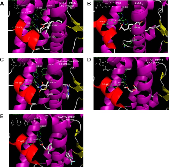
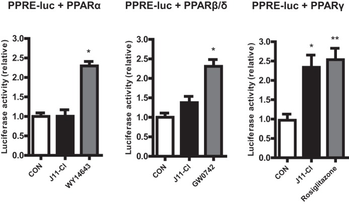
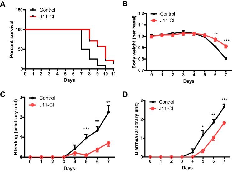
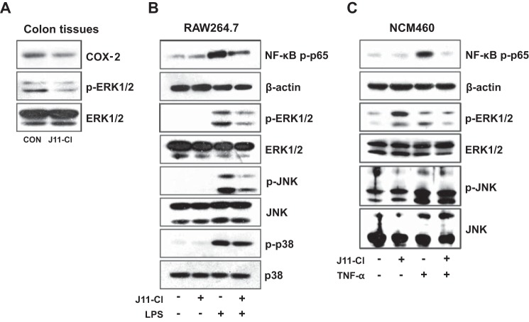
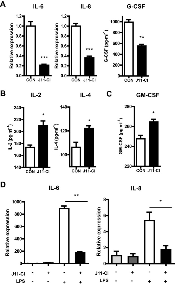
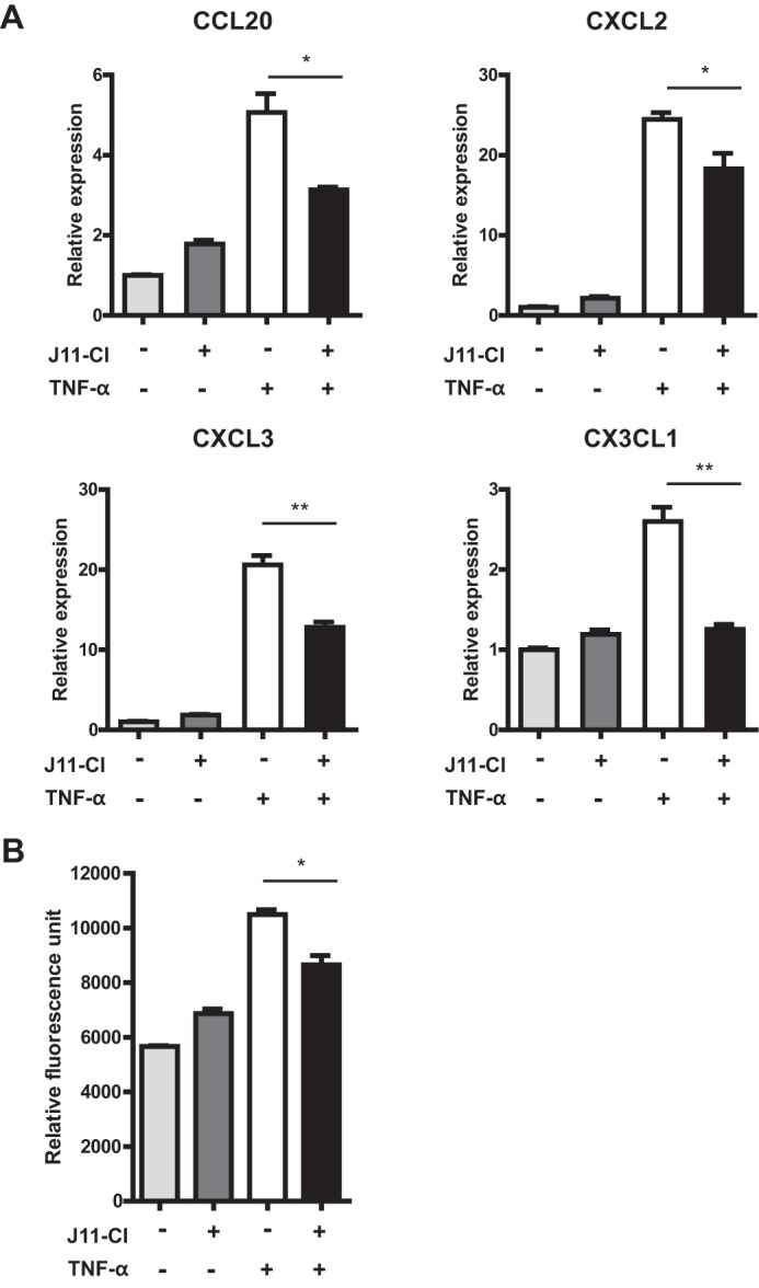
Inflammatory bowel disease (IBD) is a chronic inflammatory disease with increasing incidence and prevalence worldwide. Here we investigated the newly synthesized jasmonate analogue 2-hydroxyethyl 5-chloro-4,5-didehydrojasmonate (J11-Cl) for its anti-inflammatory effects on intestinal inflammation. First, to test whether J11-Cl can activate peroxisome proliferator-activated receptors (PPARs), we performed docking simulations because J11-Cl has a structural similarity with anti-inflammatory 15-deoxy-Δ(12,14)-prostaglandin J2 (15d-PGJ2), one of the endogenous ligands of PPARγ. J11-Cl bound to the ligand binding domain of PPARγ in the same manner as 15d-PGJ2 and rosiglitazone, and significantly increased transcriptional activity of PPARγ. In animal experiments, colitis was significantly reduced in mice with J11-Cl treatment, determined by analyses of survival rate, body weight changes, clinical symptoms, and histological evaluation. Moreover, J11-Cl decreased production of pro-inflammatory cytokines including IL-6, IL-8, and G-CSF as well as chemokines including chemokine (C-C motif) ligand (CCL)20, chemokine (C-X-C motif) ligand (CXCL)2, CXCL3, and chemokine (C-X3-C motif) ligand 1 (CX3CL1) in colon tissues, and LPS or TNF-α-stimulated macrophages and epithelial cells. In contrast, production of anti-inflammatory cytokines including IL-2 and IL-4 as well as the proliferative factor, GM-CSF, was increased by J11-Cl. Furthermore, inhibition of MAPKs and NF-κB activation by J11-Cl was also observed. J11-Cl reduced intestinal inflammation by increasing the transcriptional activity of PPARγ and modulating inflammatory signaling pathways. Therefore, our study suggests that J11-Cl may serve as a novel therapeutic agent against IBD.

Keywords: NF-kappa B (NF-κB); colitis; cytokine; inflammatory bowel disease (IBD); mitogen-activated protein kinase (MAPK); peroxisome proliferator-activated receptor (PPAR).


Figures

FIGURE 1.
FIGURE 1.
Interactions of rosiglitazone, 15d-PGJ2, GW0742, and J11-Cl with the ligand binding domain of either PPARγ or PPARβ/δ. A, docking simulation of J11-Cl to the ligand binding domain of PPARγ. B, cocrystal structure of 15d-PGJ2 with PPARγ (PDB code number 2ZK1). C, cocrystal structure of rosiglitazone with PPARγ (PDB code number 2PRG). D, docking simulation of J11-Cl to the ligand binding domain of PPARβ/δ. E, cocrystal structure of GW0742 with PPARβ/δ (PDB code number 3TKM).
FIGURE 2.
FIGURE 2.
J11-Cl increased transcriptional activity of PPARγ. Transcriptional activity of PPARγ, not PPARα and PPRAβ/δ, was significantly increased by J11-Cl. Data are shown as the mean ± S.E. A single asterisk (*) indicates p < 0.05; double asterisks (**) indicate p < 0.01.
FIGURE 3.
FIGURE 3.
J11-Cl significantly increased survival of mice and alleviated symptoms of colitis. Mice were supplied with 3% DSS in drinking water for 11 days. One group of mice was treated with J11-Cl (50 mg·kg−1) and the other group of mice was treated with vehicle (1% DMSO and 5% cremophor in saline) daily by intraperitoneal injection. The mice were monitored for rectal bleeding and diarrhea (scaled 0–4) for the duration of the experiment. A, analysis of survival rate using the log-rank test indicated that J11-Cl significantly increased survival of mice (p = 0.0013). B, body weight loss in mice treated with J11-Cl was less severe than that in the control group (vehicle treated mice). C, rectal bleeding was relieved by J11-Cl compared with the control group. D, diarrhea was relieved by J11-Cl compared with the control group. Data are shown as the mean ± S.E.; n = 12 (control group), n = 14 (J11-Cl group). A single asterisk (*) indicates p < 0.05; double asterisks (**) indicate p < 0.01; triple asterisks (***) indicate p < 0.001.
FIGURE 4.
FIGURE 4.
Tissues from J11-Cl treated mice showed less severe colonic inflammation. A, colon tissues were stained for H&E. Control mice showed more severe tissue damage with loss of crypt architecture and greater infiltration of inflammatory cells than J11-Cl-treated mice. Bar, 100 μm. B, colon tissues were stained for neutrophil surface marker, Ly-6G. Infiltration of neutrophils (brown) was significantly decreased by J11-Cl treatment. Bar, 30 μm. The number of infiltrated neutrophils was counted. C, colon tissues were stained for T cell marker, CD3. Infiltration of T cells (green) was significantly decreased by J11-Cl treatment. Arrowheads indicate CD3-positive cells. Bar, 10 μm. The number of CD3 positive cells was counted. For counting neutrophil and CD3-positive cell number, cells were counted in four randomly picked fields from two independent colon tissues in each group. Data are shown as the mean ± S.E. A single asterisk (*) indicates p < 0.05; triple asterisks (***) indicate p < 0.001.
FIGURE 5.
FIGURE 5.
J11-Cl modulated pro-inflammatory signaling pathways. A, expression of COX-2 and p-ERK1/2 in the colon tissues was determined by immunoblotting, using total ERK1/2 as an internal control. B, RAW264.7 cells were pretreated with 50 μm J11-Cl for 30 min and stimulated with 20 ng·ml−1 LPS for 15 min. Expression of phosphorylated NF-κB p65 and MAPKs (p-ERK1/2, p-JNK, and p-p38) was determined by immunoblotting, using β-actin and total MAPKs as an internal control. C, NCM460 cells were pretreated with 50 μm J11-Cl for 30 min and stimulated with 1 ng·ml−1 TNF-α for 15 min. Expression of p-NF-κB p65, p-ERK1/2, and p-JNK was determined by immunoblotting, using β-actin, ERK1/2, and JNK as an internal control.
FIGURE 6.
FIGURE 6.
J11-Cl modulated expression of pro-inflammatory, anti-inflammatory and proliferative cytokines. A, expression of pro-inflammatory cytokines in mouse colon tissues. The protein level of G-CSF and mRNA levels of IL-6 and IL-8 were decreased by J11-Cl. B, expression of anti-inflammatory cytokines in the mouse colon tissues. The protein levels of IL-2 and IL-4 were increased by J11-Cl. C, expression of proliferative cytokine in the colon tissues from mice. The protein level of GM-CSF was increased by J11-Cl. D, expression of pro-inflammatory cytokines in LPS-treated RAW264.7 cells. The cells were pretreated with 50 μm J11-Cl for 30 min and stimulated with 20 ng·ml−1 LPS for 3 h. The mRNA levels of IL-6 and IL-8 were decreased by J11-Cl. Data are shown as the mean ± S.E. A single asterisk (*) indicates p < 0.05; double asterisks (**) indicate p < 0.01; triple asterisks (***) indicate p < 0.001.
FIGURE 7.
FIGURE 7.
The mRNA expression of inflammatory chemokines in colonic epithelial cells and the number of migrated leukocytes was decreased by J11-Cl. A, NCM460 cells were pretreated with 50 μm J11-Cl for 30 min and stimulated with 1 ng·ml−1 TNF-α for 3 h. Relative gene expression of CCL20, CXCL2, CXCL3, and CX3CL1 was significantly decreased by J11-Cl treatment in TNF-α-treated NCM460 cells. B, NCM460 cells were pretreated with 50 μm J11-Cl for 30 min and stimulated with 10 ng·ml−1 TNF-α for 24 h. Relative fluorescence unit indicates migrated HL-60 cells. Data are shown as the mean ± S.E. A single asterisk (*) indicates p < 0.05; double asterisks (**) indicate p < 0.01.

References

    1. Danese S., Fiocchi C. (2011) Ulcerative colitis. NE J. Med. 365, 1713–1725 - PubMed
    1. Baumgart D. C., Sandborn W. J. (2012) Crohn's disease. Lancet 380, 1590–1605 - PubMed
    1. Molodecky N. A., Soon I. S., Rabi D. M., Ghali W. A., Ferris M., Chernoff G., Benchimol E. I., Panaccione R., Ghosh S., Barkema H. W., Kaplan G. G. (2012) Increasing incidence and prevalence of the inflammatory bowel diseases with time, based on systematic review. Gastroenterology 142, 46–54 - PubMed
    1. Shih D. Q., Targan S. R. (2009) Insights into IBD Pathogenesis. Curr. Gastroenterol. Rep. 11, 473–480 - PMC - PubMed
    1. Ananthakrishnan A. N. (2013) Environmental risk factors for inflammatory bowel disease. Gastroenterol. Hepatol. 9, 367–374 - PMC - PubMed

Publication types