Review
. 2015 Oct:34:78-86.
doi: 10.1016/j.sbi.2015.07.008. Epub 2015 Sep 2.

Insights into the structure and function of membrane-integrated processive glycosyltransferases

Affiliations
Review

Insights into the structure and function of membrane-integrated processive glycosyltransferases

Yunchen Bi et al. Curr Opin Struct Biol. 2015 Oct.

Abstract

Complex carbohydrates perform essential functions in life, including energy storage, cell signaling, protein targeting, quality control, as well as supporting cell structure and stability. Extracellular polysaccharides (EPS) represent mainly structural polymers and are found in essentially all kingdoms of life. For example, EPS are important biofilm and capsule components in bacteria, represent major constituents in cell walls of fungi, algae, arthropods and plants, and modulate the extracellular matrix in vertebrates. Different mechanisms evolved by which EPS are synthesized. Here, we review the structures and functions of membrane-integrated processive glycosyltransferases (GTs) implicated in the synthesis and secretion of chitin, alginate, hyaluronan and poly-N-acetylglucosamine (PNAG).


Conflict of interest statement

Conflict of interest statement

The authors declare no conflict of interest.

Figures

Figure 1
Figure 1
Membrane-integrated processive GTs synthesize and secrete diverse polysaccharides. The synthases may be part of multi-component complexes or function on their own. The catalytically active subunits (colored brown) share an intracellular GT and a membrane-integrated domain. Alginate consists of mannuronic (yellow) and guluronic acid (green), cellulose of glucose (beige), PNAG of NAG (gray), chitin of NAG, and HA of NAG and GA (magenta) units. A dashed circle indicates the binding site for the signaling molecule cyclic-di-GMP. Lower panel: The enzymes catalyze the transfer of a nucleotide diphosphate (NDP)-activated sugar (black hexagon) to another glycosyl unit, thereby generate NDP as a second reaction product. Among the synthases shown, HAS is the only enzyme that appears to elongate the polymer at its reducing end, thereby generating an UDP-attached polysaccharide. OM, IM: Outer and inner membrane.
Figure 2
Figure 2
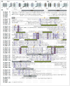
Sequence alignment and predicted secondary structure of selected family-2 GTs. (a) Predicted TM topology of Rhodobacter sphaeroides BcsA, Homo sapiens HAS2, Pseudomonas aeruginosa Alg8, Klebsiella pneumoniae PgaC, and Saccharomyces cerevisiae CHS3. Topology diagrams are shown from the N to the C terminus, labeled N and C for BcsA. GT: glycosyltransferase domain. The membrane region is shown as a blue rectangle. Topologies were predicted with TOPCONS [71]. (b) Multiple sequence alignment of the sequences used in (a). The sequences were aligned in CLUSTALW [72] and predicted TM topologies and secondary structures were used to manually refine the alignment. Predicted TM helices and secondary structure elements are shown as gray bars (TM helices) and green columns and yellow arrows for α-helices and β-strands, respectively. Conserved sequence motifs are framed with a red box and numbered 1–4.
Figure 3
Figure 3
Chemical diversity of polysaccharides. Coordinates for the shown oligosaccharides were obtained and adjusted from pdb entries 4P02 (cellulose), 3AFL (alginate), 2JCQ (HA), 4P7R (PNAG), and 3WH1 (chitin). The carbon atoms of the individual sugars are colored as in Figure 1, oxygen and nitrogen atoms are colored red and blue, respectively. The polymer’s reducing ends are labeled with an asterisk.
Figure 4
Figure 4
Pore organization of bacterial cellulose synthase subunit BcsA. BcsA’s TMHs are shown as a gray surface with the exception of TMH5 and IF2, which contains the ‘QxxRW’ motif. The GT domain is shown as a blue cartoon and cellulose and UDP are shown as sticks with blue and yellow carbon atoms, respectively. Selected residues belonging to the conserved DDG (D180), DxD (D246 and 248), TED (D343), and Q/ LxxRW (W383) motifs are shown as sticks. Cellulose synthase elongates the polymer at the non-reducing end, which is stabilized at the acceptor site within the catalytic pocket.

Similar articles

Cited by

References

    1. Lairson LL, Henrissat B, Davies GJ, Withers SG. Glycosyltransferases: structures, functions, and mechanisms. Annu Rev Biochem. 2008;77:521–555. A comprehensive review of the structures, functions and reaction mechanisms of glycosyltransferases. - PubMed
    1. McFarlane HE, Döring A, Persson S. The cell biology of cellulose synthesis. Annu Rev Plant Biol. 2014;65:69–94. - PubMed
    1. McNamara J, Morgan JLW, Zimmer J. A molecular description of cellulose biosynthesis. Annu Rev Biochem. 2015;84:17.11–17.27. - PMC - PubMed
    1. Morgan J, Strumillo J, Zimmer J. Crystallographic snapshot of cellulose synthesis and membrane translocation. Nature. 2013;493:181–186. The crystal structure of bacterial cellulose synthase provides the first insights into the mechanism of processive GTs. - PMC - PubMed
    1. Morgan JLW, McNamara JT, Zimmer J. Mechanism of activation of bacterial cellulose synthase by cyclic di-GMP. Nat Struct Mol Biol. 2014;21:489–496. The structure of cyclic-di-GMP bound to bacterial cellulose synthase reveals how cyclic-di-GMP allosterically activates cellulose biosynthesis. - PMC - PubMed

Publication types

MeSH terms