. 2015 Nov-Dec;23(6):842-54.
doi: 10.1111/wrr.12365. Epub 2015 Nov 4.

Inhibition of Pseudomonas aeruginosa biofilm formation on wound dressings

Affiliations

Inhibition of Pseudomonas aeruginosa biofilm formation on wound dressings

Kenneth S Brandenburg et al. Wound Repair Regen. 2015 Nov-Dec.

Abstract

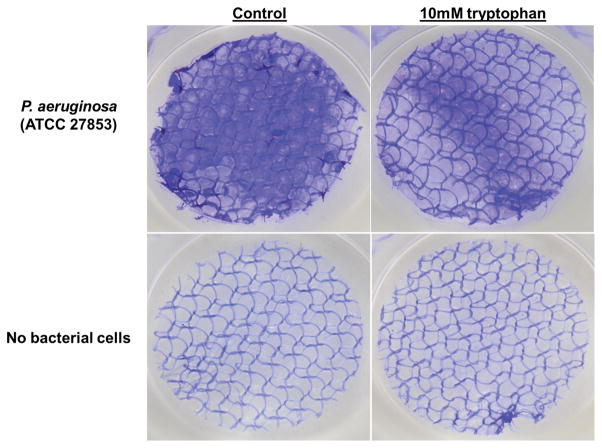
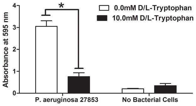
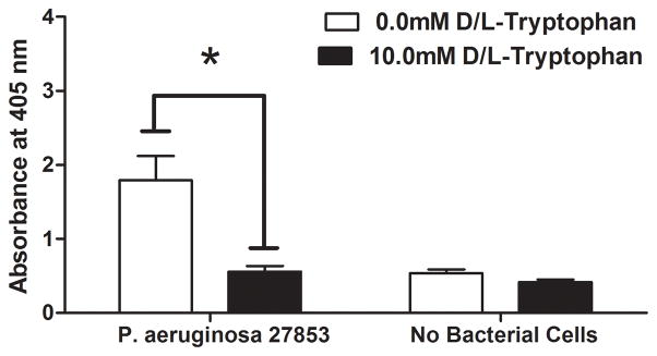
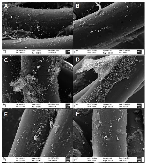
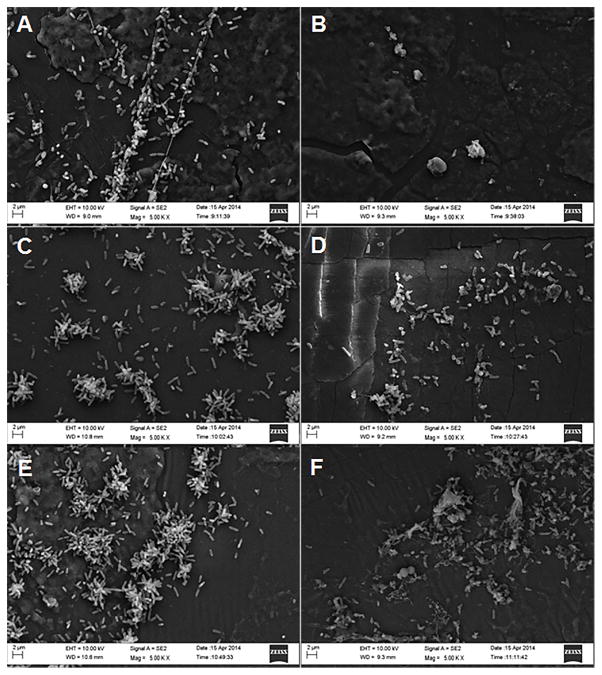
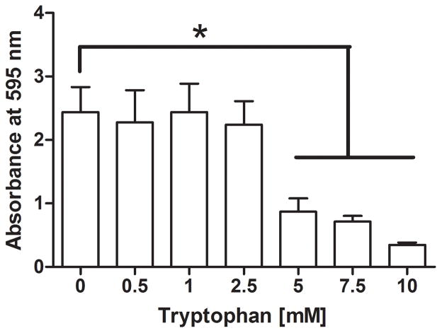
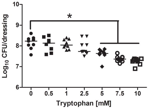
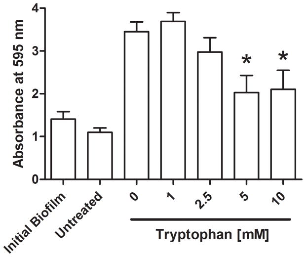
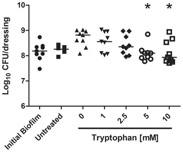
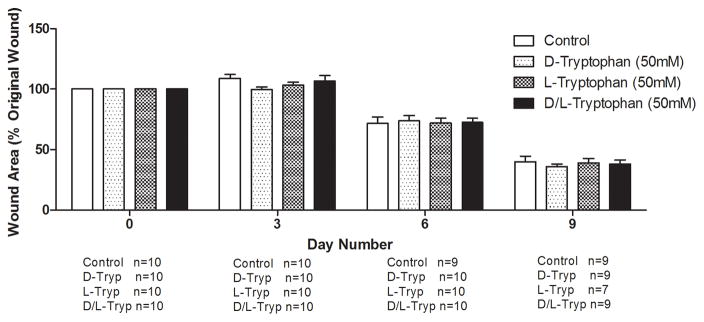
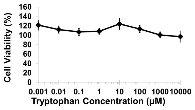
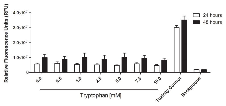
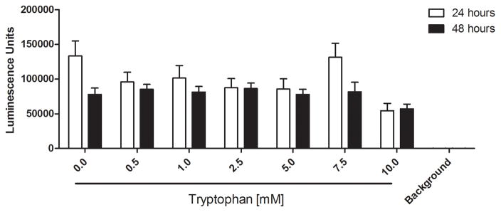
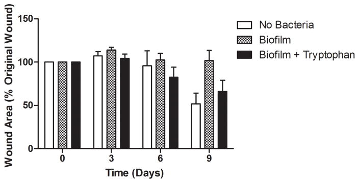
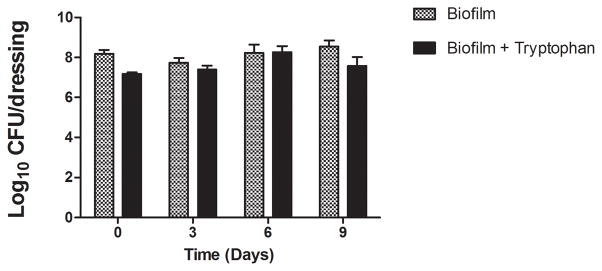
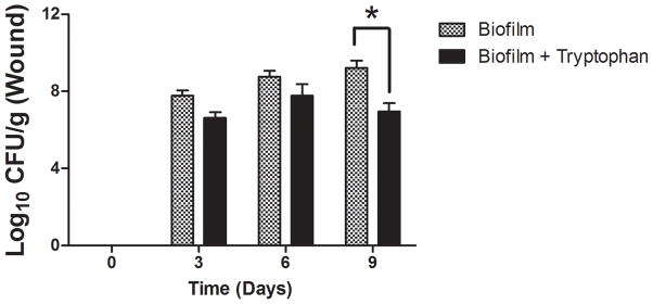
Chronic nonhealing skin wounds often contain bacterial biofilms that prevent normal wound healing and closure and present challenges to the use of conventional wound dressings. We investigated inhibition of Pseudomonas aeruginosa biofilm formation, a common pathogen of chronic skin wounds, on a commercially available biological wound dressing. Building on prior reports, we examined whether the amino acid tryptophan would inhibit P. aeruginosa biofilm formation on the three-dimensional surface of the biological dressing. Bacterial biomass and biofilm polysaccharides were quantified using crystal violet staining or an enzyme linked lectin, respectively. Bacterial cells and biofilm matrix adherent to the wound dressing were visualized through scanning electron microscopy. D-/L-tryptophan inhibited P. aeruginosa biofilm formation on the wound dressing in a dose dependent manner and was not directly cytotoxic to immortalized human keratinocytes although there was some reduction in cellular metabolism or enzymatic activity. More importantly, D-/L-tryptophan did not impair wound healing in a splinted skin wound murine model. Furthermore, wound closure was improved when D-/L-tryptophan treated wound dressing with P. aeruginosa biofilms were compared with untreated dressings. These findings indicate that tryptophan may prove useful for integration into wound dressings to inhibit biofilm formation and promote wound healing.


Figures

Figure 1
Figure 1. Tryptophan inhibits P. aeruginosa biofilm formation on a biological wound dressing
A) Representative samples of eight millimeter diameter sections of the wound dressing incubated for 48 hours at 30°C with P. aeruginosa (ATCC 27853) suspended in M63 minimal media, with or without 10mM D-/L-tryptophan. Crystal violet stained the biofilm on the dressing; samples incubated without bacterial cells were included as controls for background staining of the dressing. B) Solubilized crystal violet bound to the biofilm was quantified at a solution absorbance of 595nm. Tryptophan significantly inhibited biofilm growth on the wound dressing (*, p<0.001). C) A phosphatase linked lectin (HHA) specific for the polysaccharides of P. aeruginosa biofilms stained the biofilm matrix on the dressing. Biofilms were quantified by enzymatic cleavage of P-nitrophenylphosphate to P-nitrophenol at an absorbance of 405nm. Tryptophan significantly inhibited biofilm growth on the wound dressing (*, p=0.0032). Data is presented as the mean ± SEM of three independent experiments performed in triplicate.
Figure 2
Figure 2. Scanning electron microscopy of P. aeruginosa biofilms on collagen coated nylon fibers of a biological wound dressing
Representative scanning electron microscopy images (5000X) of control P. aeruginosa biofilms (A, C, and E), and 10mM D-/L-tryptophan inhibited biofilms (B, D, and F) grown on the wound dressing for 24 h (A and B), 48 h (C and D), and 72 h (E and F). Images were taken with a LEO 1530 scanning electron microscope.
Figure 3
Figure 3. Scanning electron microscopy images of P. aeruginosa biofilms on the silicon backing of the wound dressing
Representative scanning electron microscopic images (5000X) of control P. aeruginosa biofilms (A, C, and E) and 10.0mM D-/L-tryptophan treated biofilms (B, D, and F) grown on the wound dressing for 24 h (A and B), 48 h (C and D), and 72 h (E and F). Images were taken with a LEO 1530 scanning electron microscope.
Figure 4
Figure 4. Dose-dependent inhibition of P. aeruginosa biofilms on a biological wound dressing by tryptophan
The bioloigcal wound dressing was bathed in P. aeruginosa suspended in M63 minimal media supplemented with tryptophan (0 – 10 mM) for 48 hours at 30°C. A) Tryptophan significantly inhibited biofilm formation on the dressing at concentrations above 5mM as determined by crystal violet staining (*, p<0.05). B) Tryptophan significantly decreased bacterial colonization of the dressing at concentrations above 5mM (*, p<0.05).
Figure 5
Figure 5. Tryptophan treatment of established P. aeruginosa biofilms
P. aeruginosa biofilms were grown for 48 hours at 30°C on the biological wound dressing prior to treatment with tryptophan (0 – 10 mM). The treatment lasted for an additional 24 hours at 30°C. A) Crystal violet staining revealed that tryptophan significantly inhibited additional biofilm growth (*, p<0.05) and that concentrations above 5mM were not significantly higher than the initial biofilm. B) Quantification of bacterial cells attached to the dressing showed that tryptophan significantly reduced additional bacterial colonization compared to the fresh media alone (*, p<0.05), concentrations of tryptophan above 1mM were not significantly different than the initial bacterial load on the dressing.
Figure 6
Figure 6. Tryptophan is not cytotoxic in murine skin wounds or for immortalized human keratinocytes
A) Twenty BALB/c mice were randomized to four treatment groups. Two splinted full thickness wounds were made on the backs of each mouse, and were treated with two 8mm diameter discs of Telfa® pads soaked with 60μl of either PBS (Control), 50mM D-tryptophan, 50mM L-tryptophan, or a 50:50 combination of D- and L-tryptophan (50mM total tryptophan concentration). Treatments were applied on day 0 and reapplied on days 3 and 6. Images of the wounds were taken on days 0 (baseline), 3, 6, and 9 with a Nikon D300 camera. Image J was used to calculate the wound areas, which were normalized to the baseline (day 0) values. No significant differences in remaining wound area were measured between any treatment and the control, a p-value of below 0.05 was considered significant. B) A one hour exposure of the immortalized HaCaT cell line to D-tryptophan at 37°C exhibited no direct cytotoxicity. Cell viability was measured using calcein-AM and measuring resulting fluorescence at EM:485nm/EX:528nm. C) No loss of cellular membrane integrity was observed in immortalized human keratinocytes (STINKs) with D-/L-tryptophan concentrations up to 10mM over 24 and 48 hour incubations at 37°C using the CellTox Green Cytotoxicity Assay. D) Altered or reduced the cellular metabolism of the STINKs cell line was observed over 24 and 48 hour incubations with increasing D-/L-tryptophan concentrations as assessed by the RealTime-Glo MT Cell Viability Assay.
Figure 7
Figure 7. P. aeruginosa biofilms on wound dressings delay wound healing
P. aeruginosa biofilms with or without 10mM D-/L-tryptophan were established on wound dressings for 48 hours at 30°C. The contaminated dressings were applied to mouse skin wounds for up to 9 days. On days 3, 6, and 9, two mice from each group were sacrificed for imaging of the wounds, bacterial quantification of the dressing and wound bed. A) Representative images of the wounds in the three groups on days 0 and 9. B) Wound areas are expressed as percentages of the day 0 baseline (100%). By day 9 the control wounds without P. aeruginosa were ~50% closed, the biofilms grown in the presence of tryptophan were ~35% closed, and the biofilms grown without tryptophan remained fully open. C) Bacterial loads on the dressing (log10 CFU/dressing) did not change significantly over the 9 days on the mouse skin wounds. D) Tryptophan slightly reduced the bacterial loads on the wound bed (log10 CFU/g) with significantly lower counts detectible on day 9.

Similar articles

Cited by

References

    1. Akers KS, Mende K, Cheatle KA, Zera WC, Yu X, Beckius ML, et al. Biofilms and persistent wound infections in United States military trauma patients: a case-control analysis. BMC infectious diseases. 2014;14:190. doi: 10.1186/1471-2334-14-190. Epub 2014/04/10. - DOI - PMC - PubMed
    1. James GA, Swogger E, Wolcott R, Pulcini E, Secor P, Sestrich J, et al. Biofilms in chronic wounds. Wound repair and regeneration : official publication of the Wound Healing Society [and] the European Tissue Repair Society. 2008;16(1):37–44. doi: 10.1111/j.1524-475X.2007.00321.x. Epub 2007/12/19. - DOI - PubMed
    1. Flemming H-C, Wingender J. The biofilm matrix. Nature reviews Microbiology. 2010;8:623–33. doi: 10.1038/nrmicro2415. - DOI - PubMed
    1. López D, Vlamakis H, Kolter R. Biofilms. Cold Spring Harbor perspectives in biology. 2010;2:a000398. doi: 10.1101/cshperspect.a000398. - DOI - PMC - PubMed
    1. Ma L, Jackson KD, Landry RM, Parsek MR, Wozniak DJ. Analysis of Pseudomonas aeruginosa conditional psl variants reveals roles for the psl polysaccharide in adhesion and maintaining biofilm structure postattachment. Journal of bacteriology. 2006;188:8213–21. doi: 10.1128/JB.01202-06. - DOI - PMC - PubMed

Publication types