

Signaling Circuits and Regulation of Immune Suppression by Ovarian Tumor-Associated Macrophages
- PMID: 26343197
- PMCID: PMC4494355
- DOI: 10.3390/vaccines3020448
Signaling Circuits and Regulation of Immune Suppression by Ovarian Tumor-Associated Macrophages
Abstract
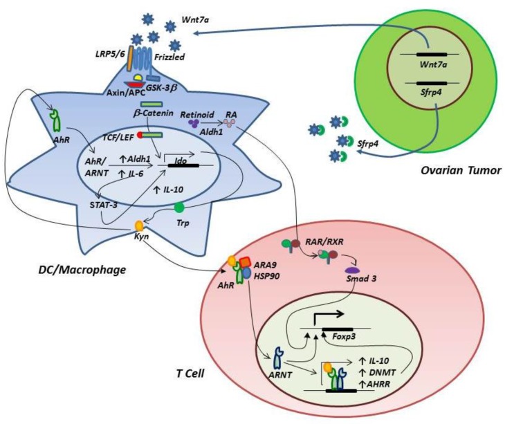
The barriers presented by immune suppression in the ovarian tumor microenvironment present one of the biggest challenges to development of successful tumor vaccine strategies for prevention of disease recurrence and progression following primary surgery and chemotherapy. New insights gained over the last decade have revealed multiple mechanisms of immune regulation, with ovarian tumor-associated macrophages/DC likely to fulfill a central role in creating a highly immunosuppressive milieu that supports disease progression and blocks anti-tumor immunity. This review provides an appraisal of some of the key signaling pathways that may contribute to immune suppression in ovarian cancer, with a particular focus on the potential involvement of the c-KIT/PI3K/AKT, wnt/β-catenin, IL-6/STAT3 and AhR signaling pathways in regulation of indoleamine 2,3-dioxygenase expression in tumor-associated macrophages. Knowledge of intercellular and intracellular circuits that shape immune suppression may afford insights for development of adjuvant treatments that alleviate immunosuppression in the tumor microenvironment and enhance the clinical efficacy of ovarian tumor vaccines.
Keywords: STAT3; aryl hydrocarbon receptor; c-KIT; dendritic cells; indoleamine 2,3-dioxygenase; macrophages; ovarian cancer; regulatory T cells.
Figures

References
-
- Curiel T.J., Coukos G., Zou L., Alvarez X., Cheng P., Mottram P., Evdemon-Hogan M., Conejo-Garcia J.R., Zhang L., Burow M., et al. Specific recruitment of regulatory T cells in ovarian carcinoma fosters immune privilege and predicts reduced survival. Nat. Med. 2004;10:942–949. doi: 10.1038/nm1093. - DOI - PubMed
-
- Wolf D., Wolf A.M., Rumpold H., Fiegl H., Zeimet A.G., Muller-Holzner E., Deibl M., Gastl G., Gunsilius E., Marth C., et al. The expression of the regulatory T cell-specific forkhead box transcription factor FoxP3 is associated with poor prognosis in ovarian cancer. Clin. Cancer Res. 2005;11:8326–8331. doi: 10.1158/1078-0432.CCR-05-1244. - DOI - PubMed
-
- Hamanishi J., Mandai M., Iwasaki M., Okazaki T., Tanaka Y., Yamaguchi K., Higuchi T., Yagi H., Takakura K., Minato N., et al. Programmed cell death 1 ligand 1 and tumor-infiltrating CD8+ T lymphocytes are prognostic factors of human ovarian cancer. Proc. Natl. Acad. Sci. USA. 2007;104:3360–3365. doi: 10.1073/pnas.0611533104. - DOI - PMC - PubMed
-
- Okamoto A., Nikaido T., Ochiai K., Takakura S., Saito M., Aoki Y., Ishii N., Yanaihara N., Yamada K., Takikawa O., et al. Indoleamine 2,3-dioxygenase serves as a marker of poor prognosis in gene expression profiles of serous ovarian cancer cells. Clin. Cancer Res. 2005;11:6030–6039. doi: 10.1158/1078-0432.CCR-04-2671. - DOI - PubMed
-
- Inaba T., Ino K., Kajiyama H., Yamamoto E., Shibata K., Nawa A., Nagasaka T., Akimoto H., Takikawa O., Kikkawa F., et al. Role of the immunosuppressive enzyme indoleamine 2,3-dioxygenase in the progression of ovarian carcinoma. Gynecol. Oncol. 2009;115:185–192. doi: 10.1016/j.ygyno.2009.07.015. - DOI - PubMed
Publication types
LinkOut - more resources
Full Text Sources
Other Literature Sources
Miscellaneous
