

Atorvastatin and sildenafil decrease vascular TGF-β levels and MMP-2 activity and ameliorate arterial remodeling in a model of renovascular hypertension
- PMID: 26343345
- PMCID: PMC4564390
- DOI: 10.1016/j.redox.2015.08.017
Atorvastatin and sildenafil decrease vascular TGF-β levels and MMP-2 activity and ameliorate arterial remodeling in a model of renovascular hypertension
Abstract
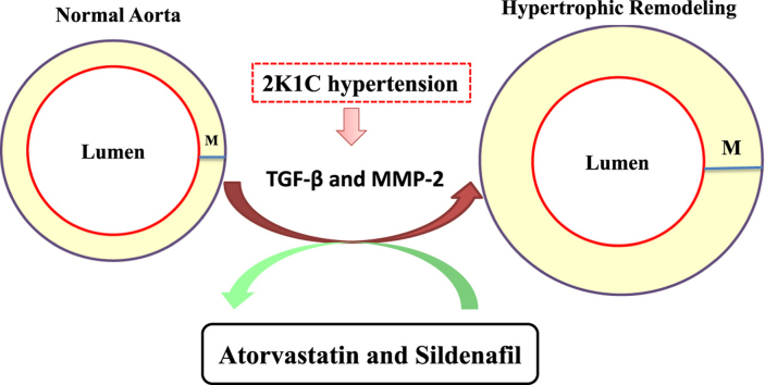
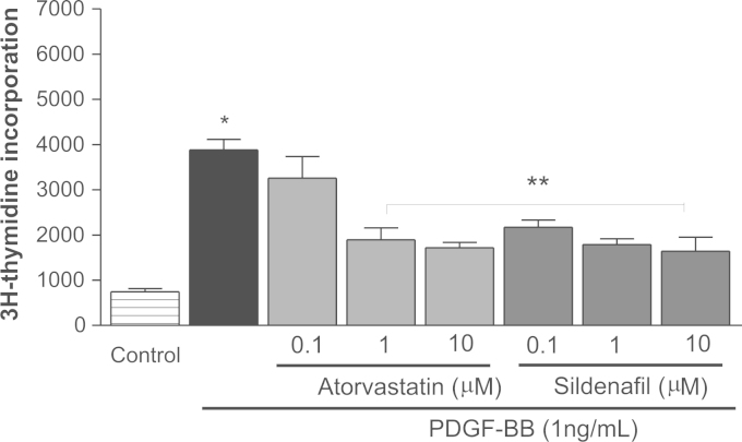
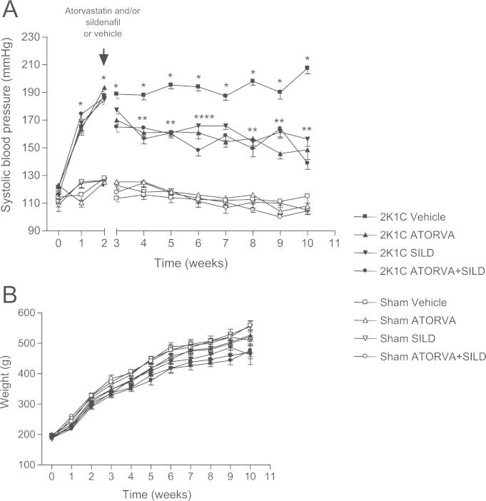
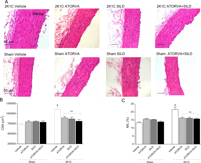
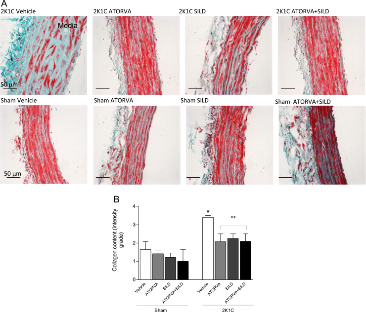
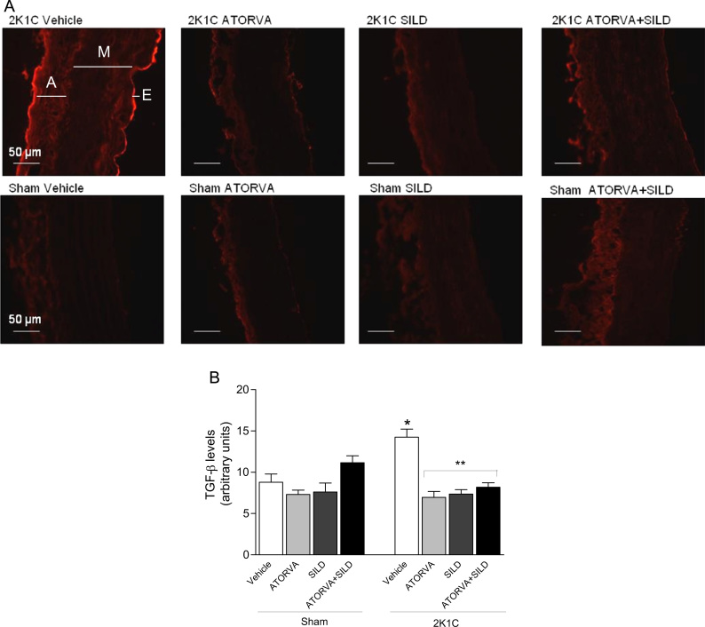
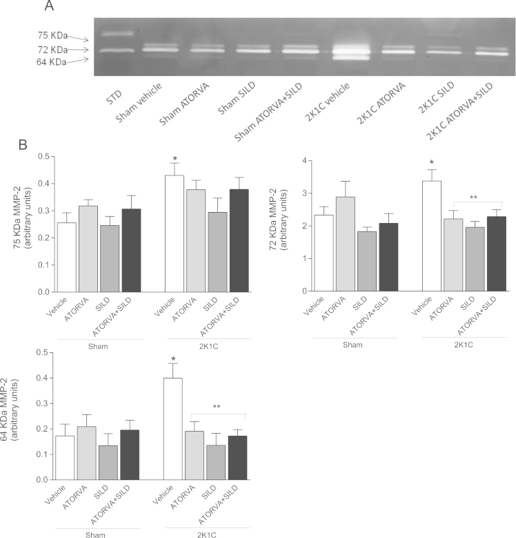
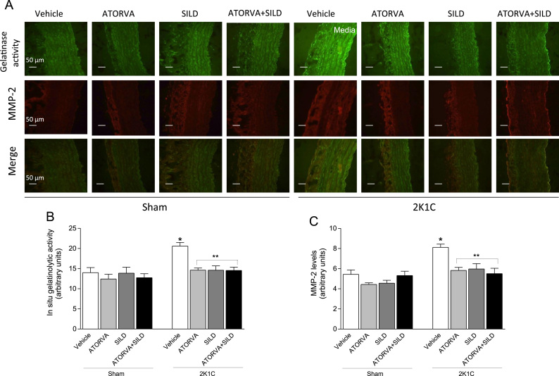
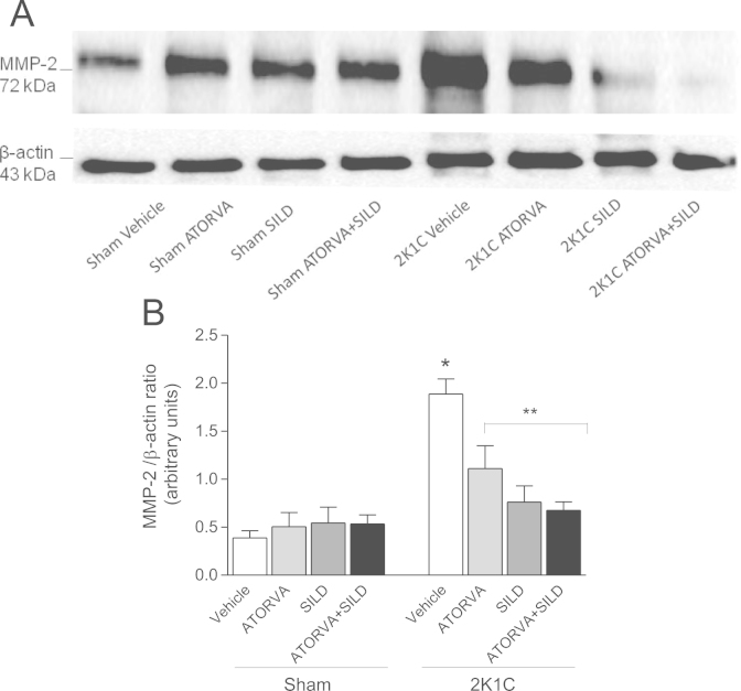
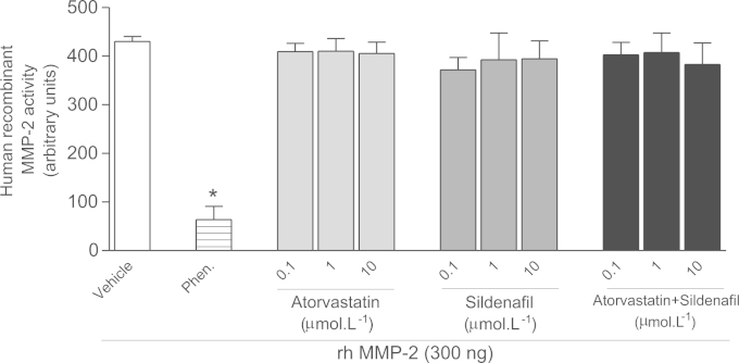
Imbalanced matrix metalloproteinase (MMP)-2 activity and transforming growth factor expression (TGF-β) are involved in vascular remodeling of hypertension. Atorvastatin and sildenafil exert antioxidant and pleiotropic effects that may result in cardiovascular protection. We hypothesized that atorvastatin and sildenafil alone or in association exert antiproliferative effects by down-regulating MMP-2 and TGF-β, thus reducing the vascular hypertrophy induced by two kidney, one clip (2K1C) hypertension. Sham and 2K1C rats were treated with oral atorvastatin 50 mg/kg, sildenafil 45 mg/kg, or both, daily for 8 weeks. Blood pressure was monitored weekly. Morphologic changes in the aortas were studied. TGF-β levels were determined by immunofluorescence. MMP-2 activity and expression were determined by in situ zymography, gel zymography, Western blotting, and immunofluorescence. The effects of both drugs on proliferative responses of aortic smooth muscle cells to PDGF and on on MMP-2 activity in vitro were determined. Atorvastatin, sildenafil, or both drugs exerted antiproliferative effects in vitro. All treatments attenuated 2K1C-induced hypertension and prevented the increases in the aortic cross-sectional area and media/lumen ratio in 2K1C rats. Aortas from 2K1C rats showed higher collagen deposition, TGF-β levels and MMP-2 activity and expression when compared with Sham-operated animals. Treatment with atorvastatin and/or sildenafil was associated with attenuation of 2K1C hypertension-induced increases in these pro-fibrotic factors. However, these drugs had no in vitro effects on hr-MMP-2 activity. Atorvastatin and sildenafil was associated with decreased vascular TGF-β levels and MMP-2 activity in renovascular hypertensive rats, thus ameliorating the vascular remodeling. These novel pleiotropic effects of both drugs may translate into protective effects in patients.
Keywords: Atorvastatin; Hypertension; Matrix metalloproteinase; Sildenafil.
Copyright © 2015 The Authors. Published by Elsevier B.V. All rights reserved.
Figures
Similar articles
-
Nitrite treatment downregulates vascular MMP-2 activity and inhibits vascular remodeling in hypertension independently of its antihypertensive effects.Free Radic Biol Med. 2019 Jan;130:234-243. doi: 10.1016/j.freeradbiomed.2018.11.002. Epub 2018 Nov 3. Free Radic Biol Med. 2019. PMID: 30399409
-
Quercetin decreases the activity of matrix metalloproteinase-2 and ameliorates vascular remodeling in renovascular hypertension.Atherosclerosis. 2018 Mar;270:146-153. doi: 10.1016/j.atherosclerosis.2018.01.031. Epub 2018 Jan 31. Atherosclerosis. 2018. PMID: 29425960
-
Temporal changes in cardiac matrix metalloproteinase activity, oxidative stress, and TGF-β in renovascular hypertension-induced cardiac hypertrophy.Exp Mol Pathol. 2013 Feb;94(1):1-9. doi: 10.1016/j.yexmp.2012.10.010. Epub 2012 Oct 13. Exp Mol Pathol. 2013. PMID: 23073243
-
Matrix Metalloproteinase 2 as a Potential Mediator of Vascular Smooth Muscle Cell Migration and Chronic Vascular Remodeling in Hypertension.J Vasc Res. 2015;52(4):221-31. doi: 10.1159/000441621. Epub 2016 Jan 6. J Vasc Res. 2015. PMID: 26731549 Review.
-
Matrix Metalloproteinases and Arterial Hypertension: Role of Oxidative Stress and Nitric Oxide in Vascular Functional and Structural Alterations.Biomolecules. 2021 Apr 16;11(4):585. doi: 10.3390/biom11040585. Biomolecules. 2021. PMID: 33923477 Free PMC article. Review.
Cited by
-
Telmisartan improves vascular remodeling through ameliorating prooxidant and profibrotic mechanisms in hypertension via the involvement of transforming growth factor-β1.Mol Med Rep. 2017 Oct;16(4):4537-4544. doi: 10.3892/mmr.2017.7177. Epub 2017 Aug 4. Mol Med Rep. 2017. PMID: 28791353 Free PMC article.
-
The Roles of Genetic Factors in Kawasaki Disease: A Systematic Review and Meta-analysis of Genetic Association Studies.Pediatr Cardiol. 2018 Feb;39(2):207-225. doi: 10.1007/s00246-017-1760-0. Epub 2017 Nov 2. Pediatr Cardiol. 2018. PMID: 29098351
-
Reductions of Circulating Nitric Oxide are Followed by Hypertension during Pregnancy and Increased Activity of Matrix Metalloproteinases-2 and -9 in Rats.Cells. 2019 Nov 7;8(11):1402. doi: 10.3390/cells8111402. Cells. 2019. PMID: 31703340 Free PMC article.
-
Sildenafil Inhibits the Growth and Epithelial-to-mesenchymal Transition of Cervical Cancer via the TGF-β1/Smad2/3 Pathway.Curr Cancer Drug Targets. 2023;23(2):145-158. doi: 10.2174/1568009622666220816114543. Curr Cancer Drug Targets. 2023. PMID: 35975844 Free PMC article.
-
Atorvastatin, Losartan and Captopril Lead to Upregulation of TGF-β, and Downregulation of IL-6 in Coronary Artery Disease and Hypertension.PLoS One. 2016 Dec 29;11(12):e0168312. doi: 10.1371/journal.pone.0168312. eCollection 2016. PLoS One. 2016. PMID: 28033321 Free PMC article.
References
-
- Sluijter J.P., de Kleijn D.P., Pasterkamp G. Vascular remodeling and protease inhibition – bench to bedside. Cardiovasc. Res. 2006;69:595–603. - PubMed
-
- Fontana V., Silva P.S., Belo V.A., Antonio R.C., Ceron C.S., Biagi C., Gerlach R.F., Tanus-Santos J.E. Consistent alterations of circulating matrix metalloproteinases levels in untreated hypertensives and in spontaneously hypertensive rats: a relevant pharmacological target. Basic Clin. Pharmacol. Toxicol. 2011;109:130–137. - PubMed
-
- Yasmin, McEniery C.M., Wallace S., Dakham Z., Pulsalkar P., Maki-Petaja K., Ashby M.J., Cockcroft J.R., Wilkinson I.B. Matrix metalloproteinase-9 (MMP-9), MMP-2, and serum elastase activity are associated with systolic hypertension and arterial stiffness. Arterioscler. Thromb. Vasc. Biol. 2005;25:372. - PubMed
-
- Castro M.M., Rizzi E., Figueiredo-Lopes L., Fernandes K., Bendhack L.M., Pitol D.L., Gerlach R.F., Tanus-Santos J.E. Metalloproteinase inhibition ameliorates hypertension and prevents vascular dysfunction and remodeling in renovascular hypertensive rats. Atherosclerosis. 2008;198:320–331. - PubMed
Publication types
MeSH terms
Substances
LinkOut - more resources
Full Text Sources
Other Literature Sources
Miscellaneous
