. 2015 Sep 14;34(5):520-31.
doi: 10.1016/j.devcel.2015.08.005. Epub 2015 Sep 3.

Co-option of an Ancestral Hox-Regulated Network Underlies a Recently Evolved Morphological Novelty

Affiliations

Co-option of an Ancestral Hox-Regulated Network Underlies a Recently Evolved Morphological Novelty

William J Glassford et al. Dev Cell. .

Abstract

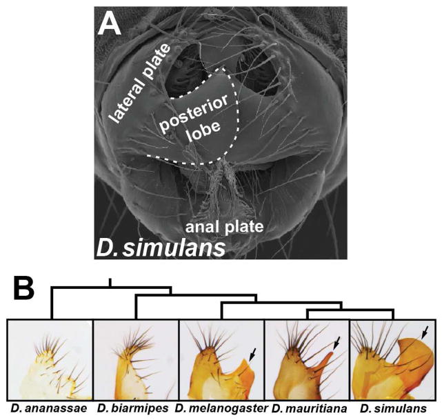
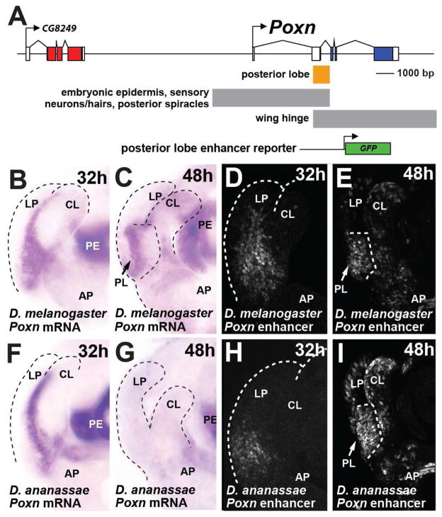
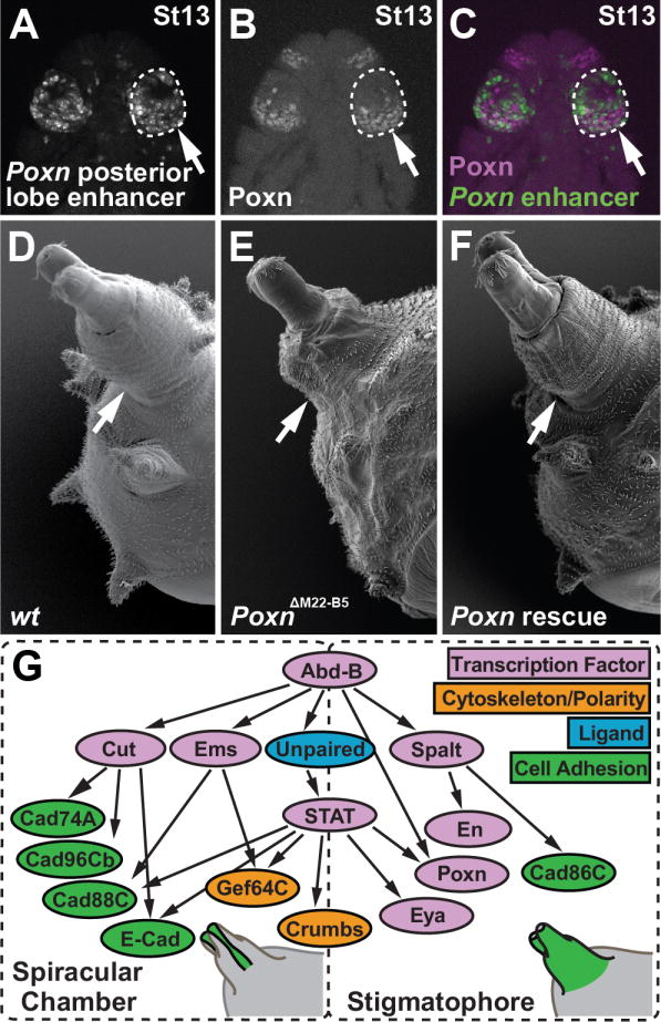
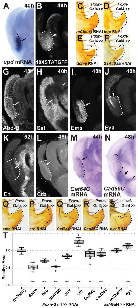
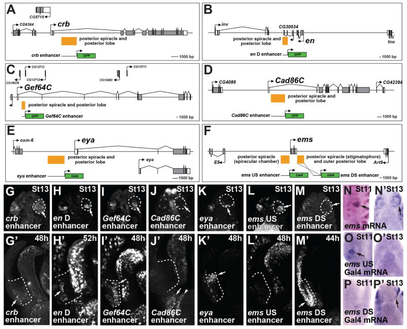
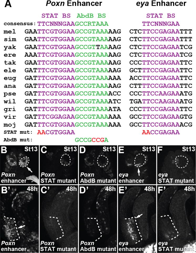
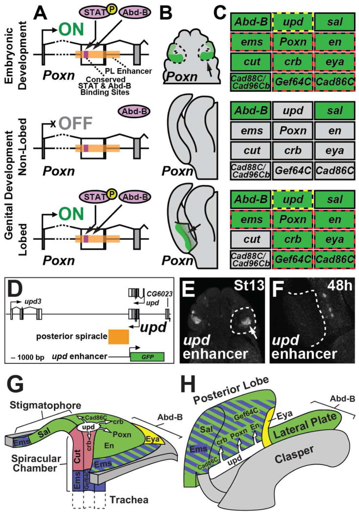
The evolutionary origins of complex morphological structures such as the vertebrate eye or insect wing remain one of the greatest mysteries of biology. Recent comparative studies of gene expression imply that new structures are not built from scratch, but rather form by co-opting preexisting gene networks. A key prediction of this model is that upstream factors within the network will activate their preexisting targets (i.e., enhancers) to form novel anatomies. Here, we show how a recently derived morphological novelty present in the genitalia of D. melanogaster employs an ancestral Hox-regulated network deployed in the embryo to generate the larval posterior spiracle. We demonstrate how transcriptional enhancers and constituent transcription factor binding sites are used in both ancestral and novel contexts. These results illustrate network co-option at the level of individual connections between regulatory genes and highlight how morphological novelty may originate through the co-option of networks controlling seemingly unrelated structures.


Figures

Figure 1
Figure 1
The posterior lobe is a morphological novelty unique to the D. melanogaster clade. (A) Scanning electron micrograph of a D. simulans male with relevant structures labeled. (B) Tree depicting the phylogenetic relationships of the species in this study, and brightfield images of their lateral plate cuticle morphologies. The posterior lobe is an outgrowth of the lateral plate unique to the melanogaster clade (arrows). See also Figure S1.
Figure 2
Figure 2
A deeply conserved enhancer of Poxn is required for posterior lobe development. (A) Schematic of the Poxn locus, displaying a subset of the described enhancer activities (Boll and Noll, 2002), and indicating the relative position of a posterior lobe reporter construct. (B, C) Accumulation of Poxn mRNA during genital development of D. melanogaster at (B) 32 hAPF and (C) 48 hAPF. (D, E) Activity of the D. melanogaster posterior lobe reporter at (D) 32 hAPF and (E) 48 hAPF. (F–G) Expression of Poxn in D. ananassae showing mRNA accumulation in the region between clasper and lateral plates (F), but not at the site where a lobe would develop (G). (H, I) Despite the absence of a posterior lobe in D. ananassae, the orthologous posterior lobe enhancer region drives expression preceding (H) and during posterior lobe development of D. melanogaster (I). CL clasper; LP lateral plate; AP anal plate, PE penis, PL posterior lobe. See also Figure S2.
Figure 3
Figure 3
The posterior lobe enhancer of Poxn is active in the Hox-regulated network of the posterior spiracle. (A) Transgenic embryo bearing the D. melanogaster posterior lobe enhancer reporter. (B) Antibody staining of Poxn protein in the posterior spiracle anlagen of the stage 13 (St13) D. melanogaster embryo presented in panel A. (C) Merged image of panels A and B, showing the Poxn enhancer (green) and Poxn protein (magenta). (D) Scanning electron micrograph of a wild-type third instar larva, showing the posterior spiracle structure. (E) The Poxn null mutant posterior spiracle is shorter relative to wildtype. (F) Rescue of posterior spiracle defects of a Poxn mutant by a fragment of the Poxn locus containing the lobe/spiracle enhancer fused to a Poxn cDNA. (G) Diagram of the posterior spiracle network, adapted from (Hu and Castelli-Gair, 1999; Lovegrove et al., 2006). The addition and placement of Poxn and eya within this network is based upon data presented in this work. Arrows in A–F point to the posterior spiracle. See also Figure S3.
Figure 4
Figure 4
Shared topology and membership of the posterior lobe and spiracle networks. Antibody staining (G–L) and in situ hybridization (A, M, N) reveal the deployment of several posterior spiracle network genes within the posterior lobe during genital development (arrows). (A–B) Expression of upd mRNA in the developing lobe (A) closely mirrors the activity of a 10XStat92E-GFP reporter (B). (C–F) Reduction in expression of members of the JAK/STAT signaling pathway hop (D), dome (E) or Stat92E (F) reduces the size of posterior lobe relative to a control (C). (G–I) The top-tier spiracle network factor Ems (I) is strongly expressed within the developing posterior lobe, while Abd-B (G) and Sal (H) are present more generally throughout the lateral plate from which the lobe emerges. (J–N) Downstream spiracle network factors Eya (J) and En (K), as well as terminal differentiation factors Crb (L), Gef64C (M), and Cad86C (N) are all expressed at specific regions and stages of posterior lobe development. (O–S) Transgenic RNAi hairpin mediated reduction in expression of spiracle network members ems (O), crb (P), Gef64C (Q), Cad86C (R), or eya (S) alters the size of posterior lobe compared to a control (shown in C). (T) Box plot depicting the relative area of posterior lobes subject to RNAi treatments, and normalized to a control. Asterisks denote significant differences (student’s paired t-test, *p <.05, ** p <.005). Dashed lines mark the position of the developing posterior lobe. (A, B, G–N) or demonstrate altered posterior lobe shape (D–F, O–S) compared to a control (C). Arrowhead in (N) identifies a pattern that is not unique to lobed species (Figure S6E). See also Figures S4 and S5.
Figure 5
Figure 5
Co-option of posterior spiracle enhancers to posterior lobe development. (A–F) Schematic diagrams of genomic loci in which an enhancer activated in both the posterior lobe and posterior spiracle were localized (orange boxes). Reporter constructs contain the schematized segment fused to either GFP or Gal4. (G-M,G′-M′) GFP reporter expression driven in transgenic D. melanogaster by enhancers for crb (G, G′), en (H, H′), Gef64C (I, I′), Cad86C (J, J′), eya(K, K′), ems US enhancer (L, L′) and ems DS enhancer (M, M′) in the posterior spiracle (G–M), and in the posterior lobe (G′-M′). (N–P) ems mRNA is first present at stage 11 in cells that contribute to the spiracular chamber, a pattern recapitulated by the ems US reporter (O), but not by the ems DS reporter (P). (N′-P′) ems is also active later during posterior spiracle development around the border of the stigmatophore (arrow) and in each embryonic segment, a pattern not encoded in the upstream enhancer (O′), but is recapitulated by the ems DS reporter (P′, arrow). (L′) The ems US reporter is not expressed within the developing posterior lobe. (J′) In addition to a lobe specific pattern (arrow), the Cad86C reporter also recapitulates a conserved pattern at the edge of the anal plate (arrowhead).
Figure 6
Figure 6
Redeployment of Poxn and eya in the posterior lobe required ancestral binding sites for Abd-B and STAT that function in the posterior spiracle context. (A) Alignment of a Stat92E binding site (purple text) and an Abd-B binding site (green text) of the Poxn lobe/spiracle enhancer and a Stat92E binding site (purple text) of the eya lobe/spiracle enhancer, showing near perfect conservation among sequenced Drosophila species. (B–D, B′-D′) Mutations to two bases in a STAT binding site (C, C′), or three bases in an Abd-B binding site (D, D′) reduces both posterior spiracle (C–D) and posterior lobe (C′-D′) activity compared to the wildtype Poxn enhancer (B, B′). Mutation of two bases in a STAT binding site (F, F′) reduces both posterior spiracle (F) and posterior lobe (F′) activity compared to the wildtype eya enhancer (E, E′). See also Figure S6.
Figure 7
Figure 7
Model depicting the co-option of genes, enhancers, and transcription factor binding sites during the origination of the novel posterior lobe. (A) (top) The posterior spiracle enhancer of Poxn binds Abd-B and phosphorylated STAT in the embryonic posterior spiracle anlagen to activate expression (“ON”). (middle) In species lacking a posterior lobe, the enhancer is not activated during genital development (“OFF”). (bottom) The deployment of regulatory factors of the spiracle network during late stages of genital development in lobed species resulted in the activation of the Poxn spiracle enhancer by Abd-B and activated STAT. (B–C) Summary of Poxn expression (B) and the status of the posterior spiracle network (C) in the three developmental contexts. (C) Expressed genes are shaded in green, while inactive genes are shaded grey. Genes activated by a shared lobe/spiracle enhancer are outlined with red dashes. The yellow dashes surrounding the upd node indicates its activation in the spiracle through an enhancer that lacks lobe activity. (D) Schematic diagram of the upd locus in which a posterior spiracle enhancer was identified (orange box). (E–F) Reporter construct containing the schematized segment fused to a GFP reporter is active in the posterior spiracle (E), but not in the posterior lobe (F). (G–H) Illustrated three-dimensional models of the developing posterior spiracle at embryonic stage 13 (G) and the developing posterior lobe (H). Important structural domains for both tissues are identified. The Hox gene Abd-B is expressed in all depicted genital structures and is deployed throughout the entire body segment containing the posterior spiracle. Zones of expression for top-tier factors Spalt (green) Ems (blue) and Cut (red) are shown. The JAK/STAT signaling pathway ligand Unpaired is shown in white, with arrows pointing toward tissues in which the JAK/STAT signaling response has been demonstrated. Downstream network genes Eya (yellow), Poxn, Engrailed, Crumbs, Gef64C and Cad86C are deployed in distinct portions of both tissues. See also Figure S7.

Comment in

Similar articles

Cited by

References

    1. Abouheif E. Establishing homology criteria for regulatory gene networks: prospects and challenges. Novartis Found Symp. 1999;222:207–221. discussion 222–225. - PubMed
    1. Bach EA, Ekas LA, Ayala-Camargo A, Flaherty MS, Lee H, Perrimon N, Baeg GH. GFP reporters detect the activation of the Drosophila JAK/STAT pathway in vivo. Gene Expr Patterns. 2007;7:323–331. - PubMed
    1. Barrio R, Shea MJ, Carulli J, Lipkow K, Gaul U, Frommer G, Schuh R, Jäckle H, Kafatos FC. The spalt-related gene of Drosophila melanogaster is a member of an ancient gene family, defined by the adjacent, region-specific homeotic gene spalt. Dev Genes Evol. 1996;206:315–325. - PubMed
    1. Bischof J, Maeda RK, Hediger M, Karch F, Basler K. An optimized transgenesis system for Drosophila using germ-line-specific phiC31 integrases. Proc Natl Acad Sci U S A. 2007;104:3312–3317. - PMC - PubMed
    1. Boll W, Noll M. The Drosophila Pox neuro gene: control of male courtship behavior and fertility as revealed by a complete dissection of all enhancers. Development. 2002;129:5667–5681. - PubMed

Publication types

MeSH terms