. 2015 Sep 14;28(3):370-83.
doi: 10.1016/j.ccell.2015.08.001. Epub 2015 Sep 3.

BRAF Mutants Evade ERK-Dependent Feedback by Different Mechanisms that Determine Their Sensitivity to Pharmacologic Inhibition

Affiliations

BRAF Mutants Evade ERK-Dependent Feedback by Different Mechanisms that Determine Their Sensitivity to Pharmacologic Inhibition

Zhan Yao et al. Cancer Cell. .

Abstract

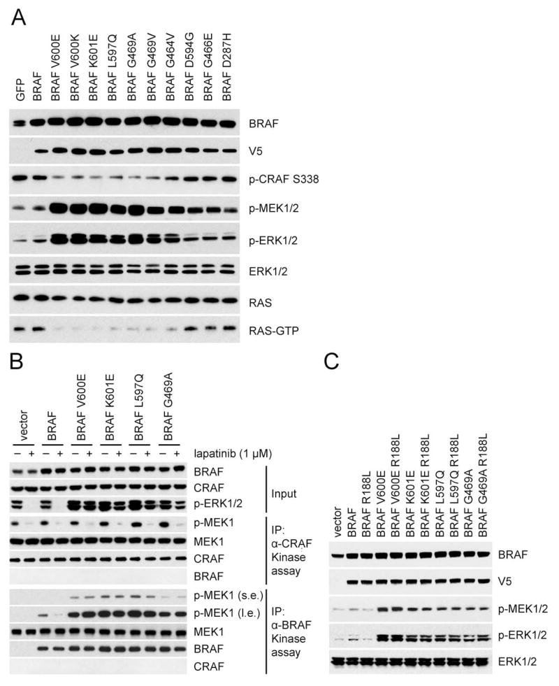
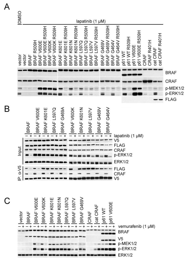
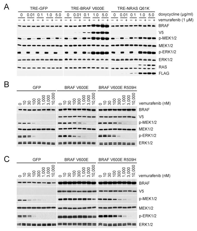
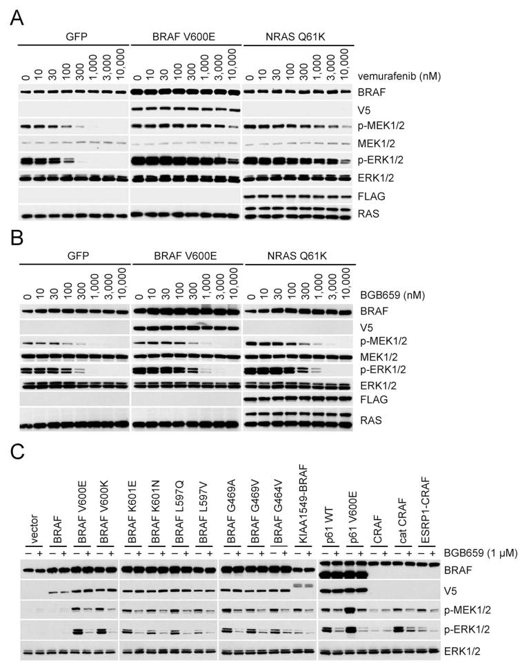
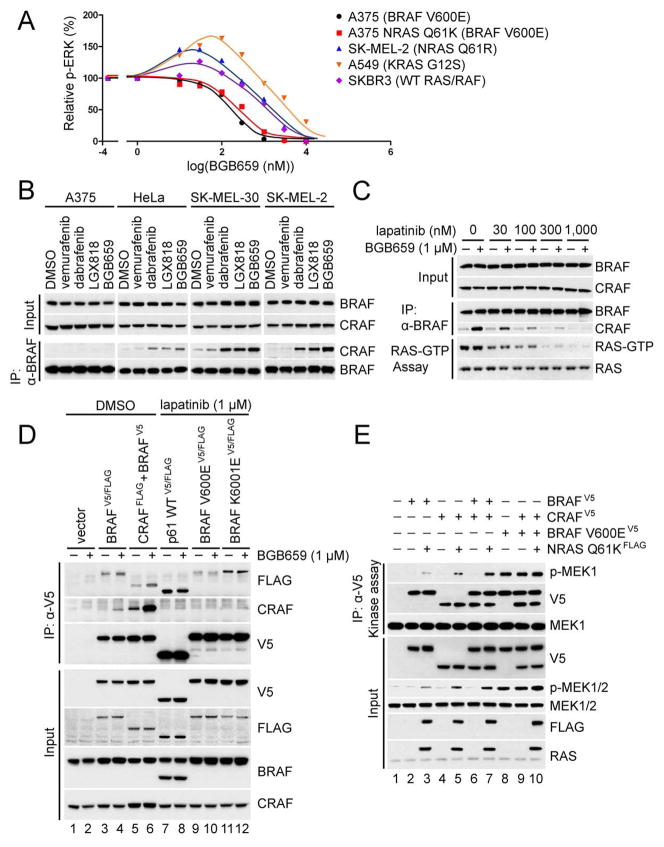
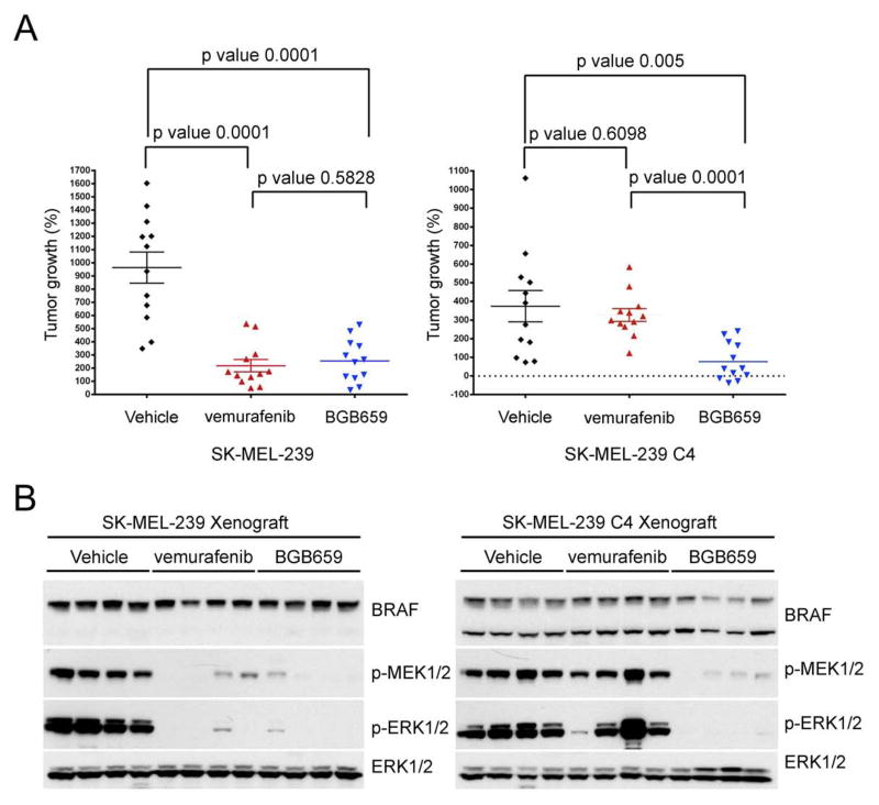
ERK signaling requires RAS-induced RAF dimerization and is limited by feedback. Activated BRAF mutants evade feedback inhibition of RAS by either of two mechanisms. BRAF V600 mutants are activated monomers when RAS activity is low; all other activating BRAF mutants function as constitutive RAS-independent dimers. RAF inhibitors effectively inhibit mutant monomers, but not dimers; their binding to one site in the dimer significantly reduces their affinity for the second. Tumors with non-V600E BRAF mutants are insensitive to these drugs, and increased expression of BRAF V600E dimers causes acquired resistance. A compound that equally inhibits both sites of mutant RAF dimers inhibits tumors driven by either class of mutants or those BRAF V600E tumors with dimer-dependent acquired resistance to monomer-specific inhibitors.


Figures

Figure 1
Figure 1. Activating BRAF mutants signal in a RAS-independent manner
(A) NIH3T3 cells stably transduced with retrovirus carrying doxycycline-inducible wild-type BRAF or the indicated mutants were treated with doxycycline (30 ng/ml) for 24 hr. Expression and/or phosphorylation of the indicated proteins were assayed by Western blot. Cellular RAS-GTP levels were determined using the active RAS pull-down assay. (B) SKBR3 cells transiently expressing V5 tagged WT BRAF or the indicated mutants were treated with lapatinib (1 μM) or DMSO for 1 hr. Cell lysate from each sample was divided into two portions for immunoprecipitation with either anti-V5 antibody or anti-CRAF antibody followed by an in vitro kinase assay with 0.5 μg K97R MEK1 protein. 5% of the whole cell extracts were used for immunoblot (Input panels). s.e.: short exposure. l.e.: longer exposure. (C) Ectopic expression of V5 tagged wild-type BRAF or the indicated mutants were expressed in SKBR3 cells followed by lapatinib treatment (1 μM, 1 hr). ERK signaling was assessed by Western blot as in Figure 1A. See also Figure S1.
Figure 2
Figure 2. Activated RAF proteins that signal as dimers are resistant to vemurafenib
(A) SKBR3 cells were transfected with the indicated plasmids. After 24 hr, the cells were treated with 1 μM lapatinib for 1 hr. Cell lysates were then analyzed by Western blot. (B) SKBR3 cells transiently co-expressing Flag-tagged and V5-tagged wild-type or mutant BRAF proteins were treated with lapatinib (1 μM, 1hr). BRAF dimerization was determined by immunoprecipitation. (C) SKBR3 cells were transfected with the indicated plasmids. After 24 hr, the cells were treated with 1 μM lapatinib for 1 hr followed by 1 μM vemurafenib for 1 hr. The indicated endogenous or ectopic proteins were assayed by Western blot. See also Figure S2.
Figure 3
Figure 3. Expression of BRAF V600E dimers causes resistance to vemurafenib
(A) A375 cells expressing inducible GFP, V5-tagged BRAF V600E or FLAG-tagged NRAS Q61K were treated with the indicated concentrations of doxycycline for 24 hr, followed by treatment with vemurafenib (1 μM, 1 hr). (B) and (C). A375 cells expressing inducible GFP, V5-tagged BRAF V600E, or V5-tagged BRAF V600E R509H were treated with either 0.02 μg/ml (B) or 0.2 μg/ml (C) doxycycline for 24 hr. Cells were then treated with vemurafenib at the indicated doses for 1 hr. Expression of the indicated proteins was assessed by Western blot. See also Figure S3.
Figure 4
Figure 4. Identification of an equipotent inhibiter of mutant BRAF monomers and dimers
(A) SKBR3 cells expressing p61 V600E or p61 V600E R509H were treated with increasing concentrations of vemurafenib or dabrafenib for 1 hr. Expression of phosphorylated MEK and phosphorylated ERK were assessed by Western blot. (B) IC50s of p-ERK inhibition for a panel of compounds in SK-MEL-239 parental cells (BRAF V600E) or the C4 clone (which expresses p61 BRAF V600E) were calculated based on densitometry analysis of Western blot results (n=3) as shown in Figure S4D. Mean values are listed in the table. (C) A375 and SK-MEL-30 cells were treated with DMSO or 3 μM LGX818 for 1hr. Cells were then washed 3 times with PBS and then placed in drug free media for the indicated times. p-ERK was assessed by Western blot (top) and quantitated by densitometry to generate the dose response curves using Prism6 (bottom); p-ERK levels relative to those from DMSO-treated cells as a function of time post treatment are shown in the accompanying graph. (D) A375, SK-MEL-239 C4 and SK-MEL-30 cells were treated with 1 μM LGX818 for 1hr followed by drug washout and treatment with the indicated compounds for an additional 1hr. The indicated fold changes of p-ERK IC50 values were calculated based on the curves as shown in Figure S4H–d. See also Figure S4 and Table S1.
Figure 5
Figure 5. BGB659 effectively inhibits vemurafenib-resistant ERK signaling
(A, B). A375 cells expressing inducible GFP(control), BRAF V600E or NRAS Q61K were treated with doxycycline (2 μg/ml, 24 hr) followed by treatment with vemurafenib (A) or BGB659 (B) at the indicated concentrations. Cell lysates were then analyzed by Western blot using the antibodies indicated. (C) SKBR3 cells were transfected with plasmids encoding the indicated wild type or mutant proteins. After 24 hr, cells were treated with BGB659 (1 μM, 1 hr). Expression and/or phosphorylation of the indicated proteins were assayed by Western blot See also Figure S5.
Figure 6
Figure 6. BGB659 preferentially inhibits signaling driven by mutant BRAF dimers
(A) The indicated cell lines were treated with 0,10,30,100, 300, 1000, 3000 and 10,000 nM BGB659 for 1 hr. Whole cell lysates were assayed by Western blot with anti-p-ERK antibody. The p-ERK level of each sample was quantitated by densitometry and then normalized to the p-ERK level in untreated cells. The p-ERK response curves were generated using Prism6. (B) A375, HeLa, SK-MEL-30 and SK-MEL-2 cells were treated with the indicated compounds for 1 hr. Endogenous BRAF was immunoprecipitated with anti-BRAF antibody. The input and isolated protein complexes were assayed by Western blot as indicated. (C) SKBR3 cells were pre-treated with lapatinib at the indicated concentrations for 1 hr followed by treatment with 1 μM BGB659 or vehicle. Cell lysates were then subjected to immunoprecipitation with anti-BRAF antibody or the RAS-GTP pull-down assay. Binding of CRAF to BRAF and RAS-GTP levels were determined by Western blot. Expression levels of BRAF and CRAF were determined using whole cell lysates. (D) The indicated RAF proteins were expressed in SKBR3 cells for 24 hr. Cells were then treated with either 1 μM lapatinib or the equivalent volume of DMSO for 1 hr, followed by treatment with 1 μM BGB659 or DMSO for an additional 1 hr. All cell lysates were collected and subjected to immunoprecipitation with an anti-V5 antibody. The isolated protein complexes and input were analyzed by Western blot. (E) BRAF-V5, CRAF-V5, BRAF V600E-V5 and FLAG tagged NRAS Q61K were transiently expressed in SKBR3 cells followed by 1hr treatment with 1 μM lapatinib. The cells were then collected and lysed, and the ectopically expressed RAF proteins were isolated with anti-V5 beads followed by elution with V5 peptide. The kinase activity of isolated RAF kinases was determined by in vitro kinase assay with K97R MEK1 as substrate. The indicated proteins from both input and kinase assays were assayed by Western blot. See also Figure S6.
Figure 7
Figure 7. BGB659 inhibits the in vivo growth of BRAF V600E tumors with acquired resistance to vemurafenib
(A) and (B) SK-MEL-239 parental or C4 clone (p61 V600E) cells were injected subcutaneously into the opposite flanks of nude mice (1.5 ×10⁷ cells per injection). After 10 days, all 12 tumors from each group were 100–150 mm3 in size and the indicated drug treatments were started The graph shows the size of each tumor after 32 days of daily treatment (A, n=12, Error bars: mean ± SEM, p values were calculated using the unpaired t test). Protein extracts from 4 random selected tumors from each group were analyzed by Western blot using the antibodies indicated (B).

Comment in

References

    1. Avraham R, Yarden Y. Feedback regulation of EGFR signalling: decision making by early and delayed loops. Nat Rev Mol Cell Biol. 2011;12:104–117. - PubMed
    1. Berghoff AS, Preusser M. BRAF alterations in brain tumours: molecular pathology and therapeutic opportunities. Curr Opin Neurol. 2014;27:689–696. - PubMed
    1. Corcoran RB, Ebi H, Turke AB, Coffee EM, Nishino M, Cogdill AP, Brown RD, Della Pelle P, Dias-Santagata D, Hung KE, et al. EGFR-mediated re-activation of MAPK signaling contributes to insensitivity of BRAF mutant colorectal cancers to RAF inhibition with vemurafenib. Cancer Discov. 2012;2:227–235. - PMC - PubMed
    1. Cutler RE, Jr, Stephens RM, Saracino MR, Morrison DK. Autoregulation of the Raf-1 serine/threonine kinase. Proc Natl Acad Sci U S A. 1998;95:9214–9219. - PMC - PubMed
    1. Davies H, Bignell GR, Cox C, Stephens P, Edkins S, Clegg S, Teague J, Woffendin H, Garnett MJ, Bottomley W, et al. Mutations of the BRAF gene in human cancer. Nature. 2002;417:949–954. - PubMed

Publication types

MeSH terms

LinkOut - more resources