. 2016 Nov;104(8):1679-1690.
doi: 10.1002/jbm.b.33515. Epub 2015 Sep 7.

Injected biodegradable polyurethane scaffolds support tissue infiltration and delay wound contraction in a porcine excisional model

Affiliations

Injected biodegradable polyurethane scaffolds support tissue infiltration and delay wound contraction in a porcine excisional model

Elizabeth J Adolph et al. J Biomed Mater Res B Appl Biomater. 2016 Nov.

Abstract

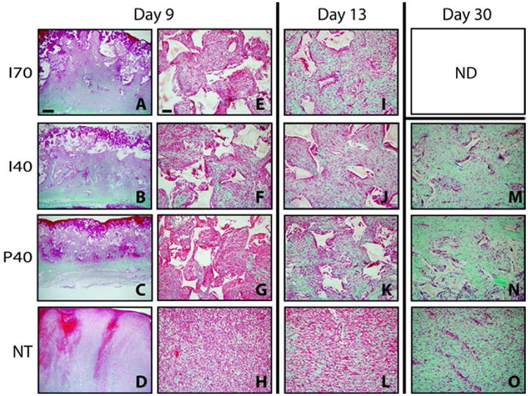
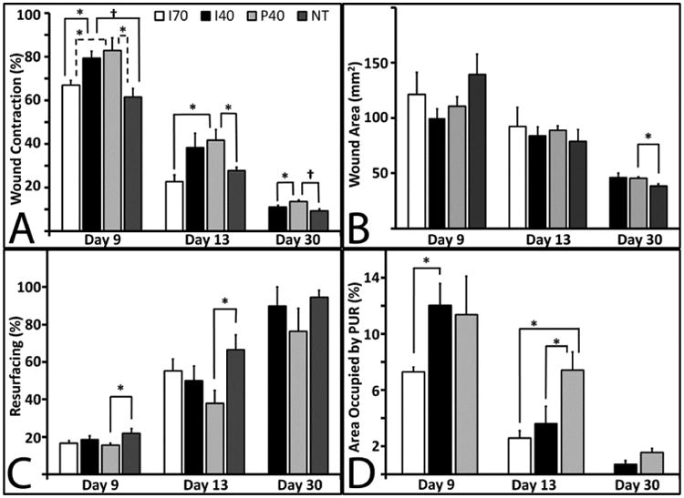
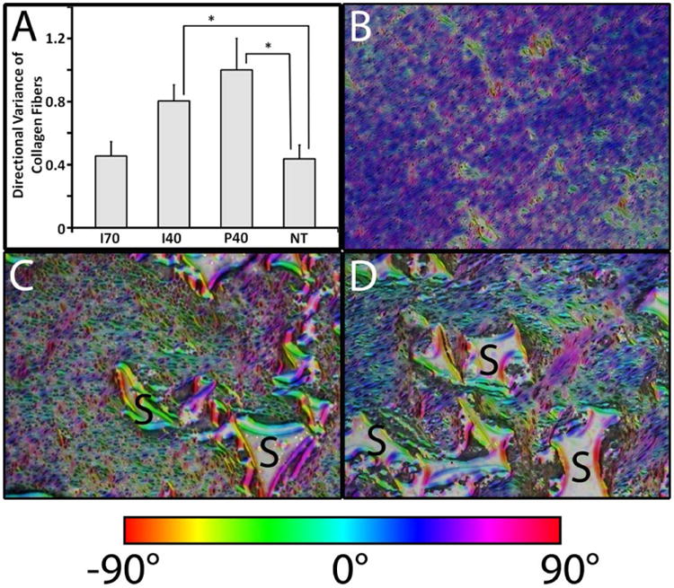
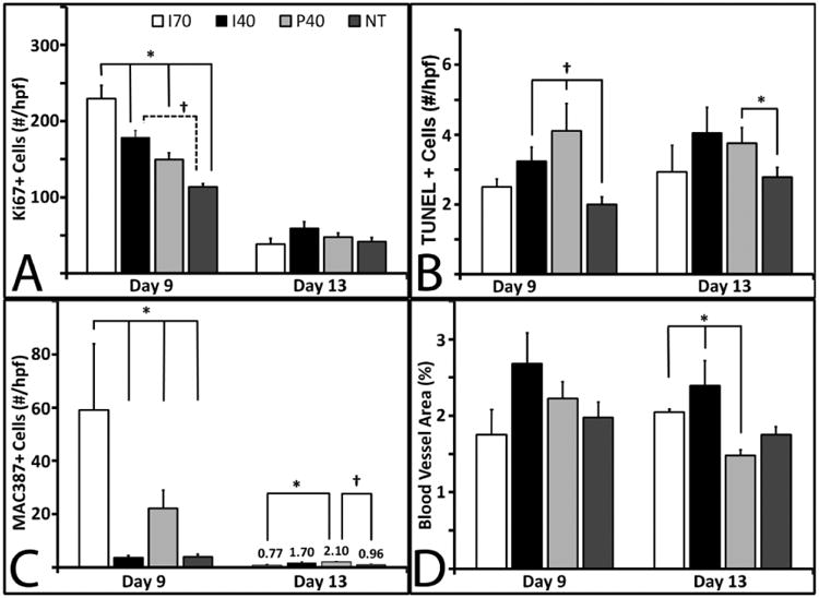
The filling of wound cavities with new tissue is a challenge. We previously reported on the physical properties and wound healing kinetics of prefabricated, gas-blown polyurethane (PUR) scaffolds in rat and porcine excisional wounds. To address the capability of this material to fill complex wound cavities, this study examined the in vitro and in vivo reparative characteristics of injected PUR scaffolds employing a sucrose porogen. Using the porcine excisional wound model, we compared reparative outcomes to both preformed and injected scaffolds as well as untreated wounds at 9, 13, and 30 days after scaffold placement. Both injected and preformed scaffolds delayed wound contraction by 19% at 9 days and 12% at 13 days compared to nontreated wounds. This stenting effect proved transient since both formulations degraded by day 30. Both types of scaffolds significantly inhibited the undesirable alignment of collagen and fibroblasts through day 13. Injected scaffolds were highly compatible with sentinel cellular events of normal wound repair cell proliferation, apoptosis, and blood vessel density. The present study provides further evidence that either injected or preformed PUR scaffolds facilitate wound healing, support tissue infiltration and matrix production, delay wound contraction, and reduce scarring in a clinically relevant animal model, which underscores their potential utility as a void-filling platform for large cutaneous defects. © 2015 Wiley Periodicals, Inc. J Biomed Mater Res Part B: Appl Biomater, 104B: 1679-1690, 2016.

Keywords: biodegradable; polyurethane scaffold; porcine model; wound contraction; wound repair.


Figures

Figure 1
Figure 1
SEM images of PUR scaffolds. Panels A and B show scaffolds constructed with 40% sucrose before (A) and after (B) leaching. Panels C and D show scaffolds constructed with 70% sucrose before (C) and after (D) leaching. Arrows indicate sucrose beads embedded in pore walls. Scale bar = 500 microns
Figure 2
Figure 2
Rheological properties of PUR scaffolds. (A) Representative cure profile of a scaffold with 40% sucrose. Vertical dashed line indicates G-crossover point, which was used to determine the working time. (B) Mean working and tack-free times of scaffolds with 40% sucrose.
Figure 3
Figure 3
Representative images of trichrome staining at day 9 with paired magnifications of 2× (left) and 20× (right). Wounds at days 13 and 30 are shown at 20× with the exception of Section M, which shows a day 30 wound injected with a scaffold with 40% sucrose at a 2× magnification. Mature collagen is intense green, nascent collagen is pale green, cytoplasm and fibrin are red or pink. PUR scaffold remnants remain unstained and appear white with angular profiles. Scale bar in A (2× magnification) = 1000 μm; Scale bar in E (20× magnification) = 100 μm.
Figure 4
Figure 4. Scaffold effects on porcine full-thickness excisional wound dimensions
For I70 at 9 and 13 days, n=4; For I40, P40, and NT, n=9 at 9 and 13 days, and n=5 at 30 days. Bars represent standard error of the mean. (A) Wound contraction measured from surface images. This graph represents the diminishing surface area as the wounds contract. A two-way ANOVA was performed to compare differences between the three time points and three treatment groups and the test of significance was met (p≤0.05). Day 9 (solid bars) and day 13 wounds (light grey bars) showed significance among the groups (p<0.05). At day 30 (dark grey), significance was noted among the groups (p<0.05). (B) Cross-sectional area of wounds. At day 30, P40 wounds were significantly larger than NT wounds (p <0.05). (C) Percent resurfacing. Resurfacing of P40 scaffolds was significantly lower than that of non-treated wounds at days 9 and 13. (D) Percent of residual PUR in the wound was significantly different at Day 13 (p=0.05) by ANOVA among the 3 different groups. At day 13, P40 levels were significantly higher than both I70 and I40 treatments (p<0.05).
Figure 5
Figure 5
Scaffold effects on the orientation of collagen fibers and cells in porcine full-thickness excisional wounds at Day 13. (A) Directional variance of alignment calculated from low-magnification images (2×) of histological sections. Significant differences (*, p< 0.05) were found between either I40 or P40 and NT treatment groups. (B-D) Representative images of Orientation J (ImageJ plug-in) generated HSB color-coded maps visualizing fiber orientation for NT (B), I40 (C), and P40 (D). Residual PUR was labeled with “S”.
Figure 6
Figure 6
Immunohistochemical measurements. Unless noted, for day 9 samples, I70, n=3; I40 and P40, n=8; and NT, n=9. For Day 13, I70, n=3; I40, n=7; and P40 and NT, n=9. Bars represent standard error of the mean. (A) Quantification of Ki67+ immunostaining at days 9 (solid bars) and 13 (grey bars). A significant difference (p ≤ 0.001) was observed among all groups at day 9. NT at day 9, n=8. (B) Quantification of TUNEL+ immunostaining at days 9 and 13. At day 9 the difference between the I40, P40, and NT groups was significant (p<0.05). (C) Quantification of MAC387+ immunostaining among the groups at days 9 and 13. Macrophage presence was significant at day 9 (p<0.001). (D) Quantification of TUNEL+ staining at days 9 and 13 only showed significance by ANOVA at Day 13 (p<0.001).

References

    1. Sen CK, Gordillo GM, Roy S, Kirsner R, Lambert L, Hunt TK, Gottrup F, Gurtner GC, Longaker MT. Human skin wounds: a major and snowballing threat to public health and the economy. Wound Repair Regen. 2009;17(6):763–71. - PMC - PubMed
    1. Zhao G, Usui ML, Lippman SI, James GA, Stewart PS, Fleckman P, Olerud JE. Biofilms and Inflammation in Chronic Wounds. Adv Wound Care (New Rochelle) 2013;2(7):389–399. - PMC - PubMed
    1. Volk SW, Iqbal SA, Bayat A. Interactions of the Extracellular Matrix and Progenitor Cells in Cutaneous Wound Healing. Adv Wound Care (New Rochelle) 2013;2(6):261–272. - PMC - PubMed
    1. Clark RAF, Ghosh K, Tonnesen MG. Tissue engineering for cutaneous wounds. Journal of Investigative Dermatology. 2007;127(5):1018–1029. - PubMed
    1. Mikos AG, Kretlow JD, Klouda L. Injectable matrices and scaffolds for drug delivery in tissue engineering. Adv Drug Deliv Rev. 2007;59(4-5):263–273. - PubMed

Publication types

LinkOut - more resources