. 2015 Nov;78(5):787-800.
doi: 10.1002/ana.24517. Epub 2015 Sep 25.

Validating novel tau positron emission tomography tracer [F-18]-AV-1451 (T807) on postmortem brain tissue

Affiliations

Validating novel tau positron emission tomography tracer [F-18]-AV-1451 (T807) on postmortem brain tissue

Marta Marquié et al. Ann Neurol. 2015 Nov.

Abstract

Objective: To examine region- and substrate-specific autoradiographic and in vitro binding patterns of positron emission tomography tracer [F-18]-AV-1451 (previously known as T807), tailored to allow in vivo detection of paired helical filament-tau-containing lesions, and to determine whether there is off-target binding to other amyloid/non-amyloid proteins.

Methods: We applied [F-18]-AV-1451 phosphor screen autoradiography, [F-18]-AV-1451 nuclear emulsion autoradiography, and [H-3]-AV-1451 in vitro binding assays to the study of postmortem samples from patients with a definite pathological diagnosis of Alzheimer disease, frontotemporal lobar degeneration-tau, frontotemporal lobar degeneration-transactive response DNA binding protein 43 (TDP-43), progressive supranuclear palsy, corticobasal degeneration, dementia with Lewy bodies, multiple system atrophy, cerebral amyloid angiopathy and elderly controls free of pathology.

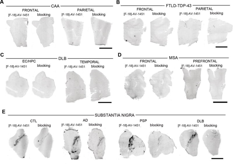
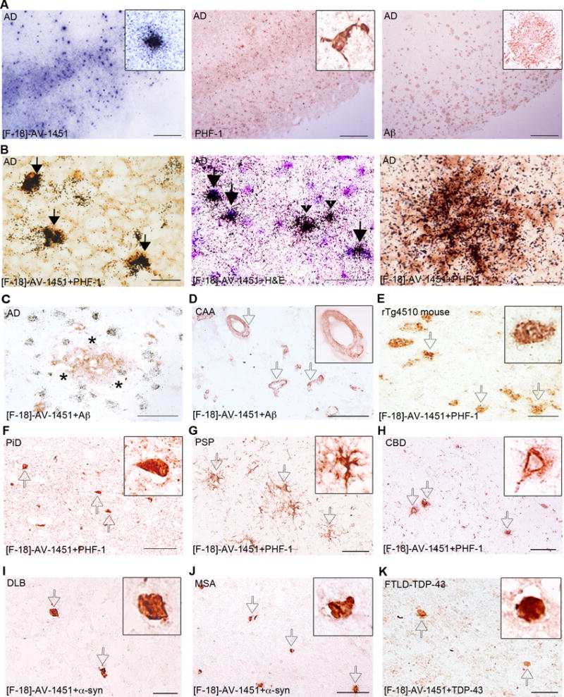
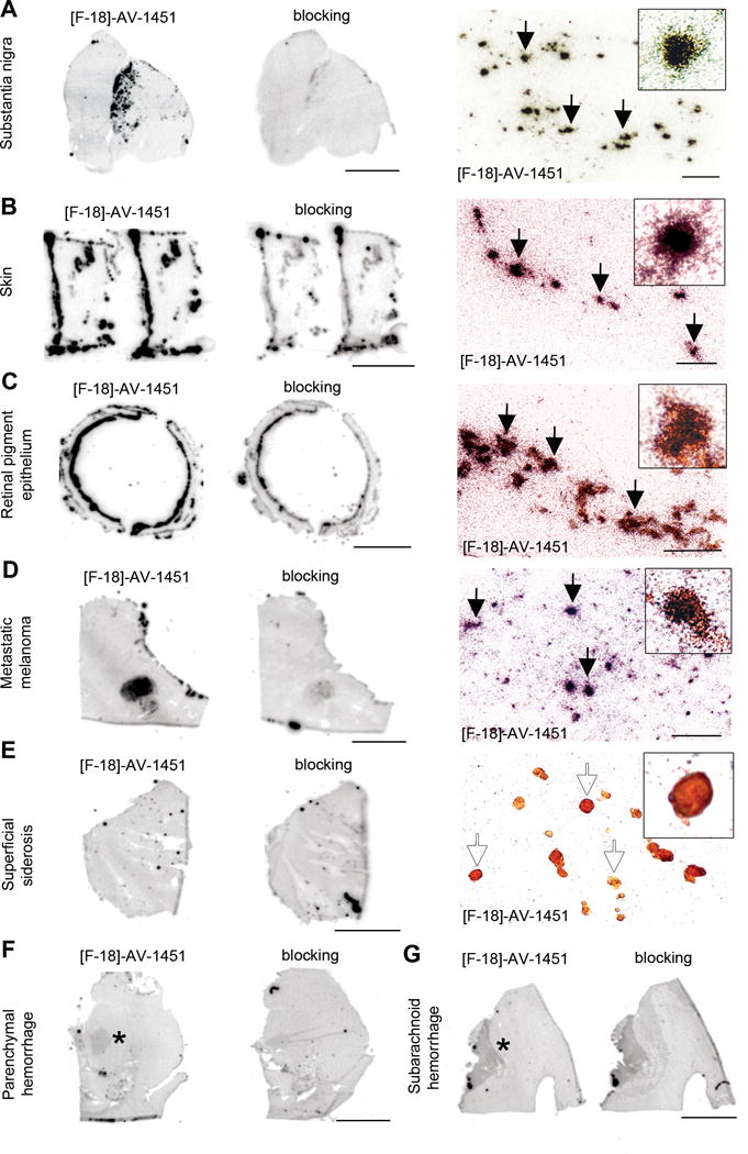
Results: Our data suggest that [F-18]-AV-1451 strongly binds to tau lesions primarily made of paired helical filaments in Alzheimer brains (eg, intraneuronal and extraneuronal tangles and dystrophic neurites), but does not seem to bind to a significant extent to neuronal and glial inclusions mainly composed of straight tau filaments in non-Alzheimer tauopathy brains or to lesions containing β-amyloid, α-synuclein, or TDP-43. [F-18]-AV-1451 off-target binding to neuromelanin- and melanin-containing cells and, to a lesser extent, to brain hemorrhagic lesions was identified.

Interpretation: Our data suggest that [F-18]-AV-1451 holds promise as a surrogate marker for the detection of brain tau pathology in the form of tangles and paired helical filament-tau-containing neurites in Alzheimer brains but also point to its relatively lower affinity for lesions primarily made of straight tau filaments in non-Alzheimer tauopathy cases and to the existence of some [F-18]-AV-1451 off-target binding. These findings provide important insights for interpreting in vivo patterns of [F-18]-AV-1451 retention.


Conflict of interest statement

Potential conflict of interest:

MM, MN, CV, IC, EB, LR, WK, CM, MI, MD, BD, SG, JG, MP, TGI have no conflicts of interest.

NV has a patent called “Improved Radiosynthesis of The Tau Imaging Radiopharmaceutical [18F]AV-1451” (PCT/US2014/040165) pending. AV-1451 is the PET compound used in this study.

KJ reports grants from Eli Lilly and Company during the conduct of the study. Eli Lilly and Company owns Avid, the company that created AV-1451, the PET compound used in this study.

BH reports personal fees from Eli Lilly and Company outside the submitted work. Eli Lilly and Company owns Avid, the company that created AV-1451, the PET compound used in this study.

Figures

Figure 1
Figure 1
[F-18]-AV-1451 phosphor screen images of brain slices from an AD case (A), a control (B), a PiD case (C), a PSP case (D), a CBD case (E) and a rTg4510 mouse brain (F). A strong [F-18]-AV-1451 binding was observed in cortical regions containing tangles and PHF-neurites from AD brains; the signal was blocked by adding unlabeled AV-1451 (A) and no [F-18]-AV-1451 signal was detected in the cerebellum (A). Slices from a control case free of pathology did not show detectable [F-18]-AV-1451 binding (B). [F-18]-AV-1451 binding was not detectable either in non-PHF tau-containing slices from a PiD case (C), a PSP case (D), a CBD case (E) and an rTg4510 mouse brain (F). Note that [F-18]-AV-1451 binding was confined to incidental age-related PHF-tangle pathology in superficial layers of EC in a PSP case (asterisk, D). Abbreviations: AD = Alzheimer’s disease; CBD = corticobasal degeneration, CTL = control, EC = entorhinal cortex, HPC = hippocampus, NFT = neurofibrillary tangles; PiD = Pick’s disease, PHF = paired helical filaments, PSP = progressive supranuclear palsy. Scale bar= 1 cm.
Figure 2
Figure 2
[F-18]-AV-1451 phosphor screen images of brain slices from a CAA carrier of the D23N Iowa APP mutation (A), a FTLD-TDP-43 case (B), a DLB case (C), a MSA case (D) and midbrain containing slices from control, AD, PSP and DLB cases (E). No [F-18]-AV-1451 binding was detected in slices containing CAA lesions (A), TDP-43 inclusions (B), Lewy bodies (C) and glial α-synuclein inclusions (D). Strong [F-18]-AV-1451 signal was observed in the region corresponding to the substantia nigra in all cases studied regardless their pathological diagnosis. (E). Abbreviations: AD = Alzheimer’s disease, APP = amyloid precursor protein, CAA = cerebral amyloid angiopathy, CTL = control, DLB = dementia with Lewy bodies, FTLD = frontotemporal lobar degeneration, MSA = multiple system atrophy, PSP = progressive supranuclear palsy, TDP-43 = TAR DNA binding protein 43. Scale bar= 1 cm.
Figure 3
Figure 3
[F-18]-AV-1451 nuclear emulsion autoradiography photomicrographs of brain slices from an AD case (A–C), a CAA carrier of the D23N Iowa APP mutation (D), a rTg4510 mouse (E), a PiD case (F), a PSP case (G) a CBD case (H), a DLB case (I), a MSA case (J) and a FTDL-TDP-43 case (K). Combined [F-18]-AV-1451 nuclear emulsion autoradiography followed by immunostaining with appropriate antibodies (B right and left panels, C–K) or H&E counter staining (B middle panel). The primary antibodies used were PHF-1 (kind gift of Dr. Peter Davies, A middle panel, B left and right panels, and E–H), anti-Aβ (A right panel, C and D), anti α-synuclein (I and J) and anti-TDP-43 (K). [F-18]-AV-1451 nuclear emulsion autoradiography showed a strong cortical accumulation of silver grains in cortical layers III and V in AD brains (A, left panel) mirroring the pattern on adjacent slices of tau PHF-1 immunostaining (A, middle panel) but not the Aβ immunostaining (A, right panel). Accumulation of silver grains from the nuclear emulsion colocalized with PHF-1 stained tangles (solid arrows, B, left panel), both intraneuronal (solid arrows) and extraneuronal (black arrowheads, B middle panel) and PHF-tau containing neurites surrounding plaques in AD brains (empty arrows, B, right panel). No detectable accumulations of silver grains were observed in association with diffuse plaques in AD (asterisk, C), amyloid-containing vessels in CAA (empty arrows, D), tangles in Tg4510 mouse brains (empty arrows, E), Pick bodies in a PiD case (empty arrows, F), astrocytic plaques in a PSP case (empty arrows, G) or coiled bodies and globose tangles in a CBD case (empty arrows, H). No silver grains were detected in association with Lewy bodies in a DLB case (empty arrows, I), α-synuclein inclusions in a MSA case (empty arrows, J) or TDP-43 inclusions in a FTLD-TDP-43 case (empty arrows, K). Abbreviations: Aβ = amyloid-β, AD = Alzheimer’s disease; APP = amyloid precursor protein, CAA = cerebral amyloid angiopathy, CBD = corticobasal degeneration, DLB = dementia with Lewy bodies, FTLD = frontotemporal lobar degeneration, H&E = hematoxylin and eosin, MSA = multiple system atrophy, PHF = paired helical filament, PiD = Pick’s disease, PSP = progressive supranuclear palsy, TDP-43 = TAR DNA binding protein 43. Scale bar = 2 mm (A), 20 μm (B), 50 μm (C–K).
Figure 4
Figure 4
[F-18]-AV-1451 phosphor screen and nuclear emulsion autoradiography images of slices containing substantia nigra in a PSP case (A), skin (B) and retinal pigment epithelium (C) in an AD case, metastatic melanoma (D), cerebellum in a superficial siderosis case (E), and parenchymal (F) and subarachnoid (G) hemorrhagic lesions. [F-18]-AV-1451 phosphor screen autoradiography images are displayed in A–E, left and middle panels, and F–G. [F-18]-AV-1451 nuclear emulsion autoradiography images are displayed in A–E, right panel. Strong [F-18]-AV-1451 binding was observed in neuromelanin-containing neurons of the substantia nigra (A), skin melanocytes (B), neuromelanin containing granules in the retinal pigment epithelium (C) and malignant melanocytes from a metastatic melanoma (D). No detectable [F-18]-AV-1451 binding was observed in association with hemosiderin deposits in a superficial siderosis case (E). Mild [F-18]-AV-1451 binding was noticed in association with intraparenchymal (asterisk, F) and subarachnoid (asterisk, G) hemorrhagic lesions. Abbreviations: AD = Alzheimer’s disease, PSP = progressive supranuclear palsy. Scale bar: 1cm (phosphor screen autoradiographic images), 50 μm (A, C, D right panels), 25 μm (B, E, right panels).

References

    1. Xia CF, Arteaga J, Chen G, et al. [(18)F] T807, a novel tau positron emission tomography imaging agent for Alzheimer’s disease. Alzheimer’s & dementia: the journal of the Alzheimer’s Association. 2013 Nov;9(6):666–76. - PubMed
    1. Zhang W, Arteaga J, Cashion DK, et al. A highly selective and specific PET tracer for imaging of tau pathologies. Journal of Alzheimer’s disease: JAD. 2012;31(3):601–12. - PubMed
    1. Chien DT, Bahri S, Szardenings AK, et al. Early clinical PET imaging results with the novel PHF-tau radioligand [F-18]-T807. Journal of Alzheimer’s disease: JAD. 2013 Jan 1;34(2):457–68. - PubMed
    1. Cairns NJ, Bigio EH, Mackenzie IR, et al. Neuropathologic diagnostic and nosologic criteria for frontotemporal lobar degeneration: consensus of the Consortium for Frontotemporal Lobar Degeneration. Acta neuropathologica. 2007 Jul;114(1):5–22. - PMC - PubMed
    1. Hauw JJ, Daniel SE, Dickson D, et al. Preliminary NINDS neuropathologic criteria for Steele-Richardson-Olszewski syndrome (progressive supranuclear palsy) Neurology. 1994 Nov;44(11):2015–9. - PubMed

Publication types

MeSH terms