. 2015 Dec:72:74-89.
doi: 10.1016/j.biomaterials.2015.08.048. Epub 2015 Aug 29.

Hyaluronic acid-decorated dual responsive nanoparticles of Pluronic F127, PLGA, and chitosan for targeted co-delivery of doxorubicin and irinotecan to eliminate cancer stem-like cells

Affiliations

Hyaluronic acid-decorated dual responsive nanoparticles of Pluronic F127, PLGA, and chitosan for targeted co-delivery of doxorubicin and irinotecan to eliminate cancer stem-like cells

Hai Wang et al. Biomaterials. 2015 Dec.

Abstract

Dual responsive nanoparticles are developed for co-delivery of multiple anticancer drugs to target the drug resistance mechanisms of cancer stem-like cells (CSCs). The nanoparticles consist of four polymers approved by the Food and Drug Administration (FDA) for medical use: Poly(d,l-lactide-co-glycolide) (PLGA), Pluronic F127 (PF127), chitosan, and hyaluronic acid (HA). By combining PLGA and PF127 together, more stable and uniform-sized nanoparticles can be obtained than using PLGA or PF127 alone. The HA is used for not only actively targeting CSCs to reduce their drug resistance due to dormancy (i.e., slow metabolism), but also replacing the commonly used poly(vinyl alcohol) as a stabilizing agent to synthesize the nanoparticles using the double-emulsion approach and to allow for acidic pH-triggered drug release and thermal responsiveness. Besides minimizing drug efflux from CSCs, the nanoparticles encapsulated with doxorubicin hydrochloride (DOX, hydrophilic) and irinotecan (CPT, hydrophobic) to inhibit the activity of topoisomerases II and I, respectively, can fight against the CSC drug resistance associated with their enhanced DNA repair and anti-apoptosis. Ultimately, the two drugs-laden nanoparticles can be used to efficiently destroy the CSCs both in vitro and in vivo with up to ∼500 times of enhancement compared to the simple mixture of the two drugs.

Keywords: CD44; Cancer stem-like cell; Co-delivery; Drug resistance; Topoisomerase.


Figures

Fig. 1
Fig. 1
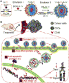
A schematic illustration of the procedure for preparing nanoparticles and their capability of targeting four different drug resistant mechanisms of cancer stem-like cells (CSCs). (A) Hyaluronic acid (HA), PF127 (PF, with and without chitosan modification), and PLGA (P) were used to prepare the DOX (D) and CPT (C)-laden HAC-PFP-DC nanoparticles using an improved double-emulsion method. (B) The HAC-PFP-DC nanoparticles can be used to fight against the multifaceted mechanisms of drug resistance of CSCs including their capability of actively targeting CSCs to facilitate drug uptake by the cells to reduce their drug resistance due to dormancy or slow metabolic activity (1); delivery and release of drugs away from the plasma membrane to minimize the drug efflux by the transmembrane pumps of CSCs (2); and co-delivery of two chemotherapeutic drugs (DOX and CPT) that synergistically inhibit the activity of topoisomerases I and II that are crucial for DNA repair and production of anti-apoptotic proteins to combat against the drug resistance of CSCs due to their enhanced capability of DNA repair (3) and anti-apoptosis (4). O: oil (i.e., organic solvent), W: water, DOX: doxorubicin hydrochloride, CPT: irinotecan, PLGA: poly (D,L-lactide-co-glycolide), and Topo: topoisomerase.
Fig. 2
Fig. 2
Nanoparticle characterization showing the uniform size, core-shell morphology, high aqueous solubility, and thermal responsiveness of the HAC-PFP nanoparticles. (A) SEM images of HAC-PFP, PLGA, and PF127 nanoparticles. (B) TEM images of HAC-PFP nanoparticles after loading with DOX and CPT (i.e., HAC-PFP-DC nanoparticles) at two different magnifications. (C) A typical picture of PLGA, HAC-PFP, HAC-PFP-DC, and PF127 nanoparticles in water where the two samples in the 1.5 ml centrifuge tubes are after centrifugation. (D) Size distribution of HAC-PFP, HAC-PFP-D, and HAC-PFP-DC nanoparticles determined by dynamic light scattering (DLS) at 22 and 37 °C. (E) Surface zeta potential of HAC-PFP, HAC-PFP-D, and HAC-PFP-DC nanoparticles synthesized with different percentages of HA at 22 °C and 37 °C. Collectively, the data show the round and core-shell morphology of the HAC-PFP nanoparticles (with or without drugs) together with their several advantages to the nanoparticles made of PLGA or PF127 alone including uniform size, convenient collection by centrifugation, and thermal responsiveness in size and surface change.
Fig. 3
Fig. 3
Characterization of drug encapsulation and release showing the acidic pH-triggered drug release from the HAC-PFP-DC nanoparticles. Normalized excitation (A) and fluorescence emission (B) spectra of HAC-PFP-DC nanoparticles showing the presence of both DOX and CPT in the nanoparticles. (C) UV-Vis absorbance of HAC-PFP-DC nanoparticles showing the presence of both DOX and CPT in the nanoparticles. (D) Fluorescence images of the HAC-PFP-DC nanoparticles before rotary evaporation to remove organic solvent showing that both DOX (red) and CPT (blue) existed in each particle. (E) In vitro release of CPT and DOX from PFP-DC and HAC-PFP-DC nanoparticles at pH 5.0 and 7.4 showing the acidic pH-triggered drug release from both (particularly the HAC-PFP-DC) nanoparticles. TEM images (F) and DLS data (G) of PFP-DC and HAC-PFP-DC nanoparticles after incubated at pH 7.4 and 5 at 37 °C for 3 h. The arrows in the TEM images of HAC-PFP-DC nanoparticles indicate compromised nanoparticles.
Fig. 4
Fig. 4
In vitro cell uptake data showing the HA coating on HAC-PFP-DC nanoparticles significantly improves drug delivery into prostasphere and mammosphere cells enriched with CSCs. Fluorescence micrographs of (A) prostasphere and (B) mammosphere cells after incubated with the simple mixture of free DOX&CPT, PFP-DC nanoparticles, and HAC-PFP-DC nanoparticles for 3 h at 37 °C. (C) Typical flow cytometry data of fluorescence intensity of DOX and CPT in both prostasphere and mammosphere cells after the three different treatments. Collectively, the data show enhanced cellular uptake of both anticancer drugs delivered using HA-PPF-DC nanoparticles via endocytosis by both mammosphere and prostasphere cells, compared to PFP-DC nanoparticles and free anticancer drugs. The LysoTracker green stains late endosomes and lysosomes.
Fig. 5
Fig. 5
The HAC-PFP-DC nanoparticles exhibit excellent anti-CSC capability in vitro. (A) Viability of prostasphere and mammosphere cells after treated with free DOX, simple mixture of free DOX&CPT, PFP-DC nanoparticles, HAC-PFP-D nanoparticles, and HAC-PFP-DC nanoparticles. (B) IC50 (inhibitory concentration to kill 50% cells) and (C) IC85 (inhibitory concentration to kill 85% cells) calculated from the viability data for the various drug formulations. (D) Growth (area normalized to that of the first day) curve of prostaspheres and mammospheres after treated with 1.72 μM drug in various formulations together with no treatment (NT) control. (E) Typical micrographs showing morphology of the prostasphere and mammospheres after treated with 1.72 μM drug in various formulations for 10 days together with no treatment (NT) control. *: p < 0.05.
Fig. 6
Fig. 6
The HAC-PFP-DC nanoparticles exhibit excellent in vivo anti-tumor capacity. (A) Typical photographs showing the size of tumors (indicated by arrows) in mice on day 30 after six different treatments. (B) Tumor growth curves for various drug formulation treatments. *: p < 0.05. (C) Weight of the tumors together with images of tumors collected after sacrificing the mice on day 30. *: p < 0.05. (D) Representative histology (H&E) images of the tumors collected on day 30. (E) Staining of early and late apoptosis of cells in the tumors collected on day 30 with Annexin V and PI, respectively. The DAPI stains cell nuclei. (F) Relative (to saline control) body weight of the mice with the various treatments showing no significant difference between the different treatments.
Fig. 7
Fig. 7
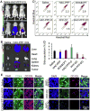
The HAC-PFP-DC nanoparticles target tumors and effectively kill CSCs in vivo. (A) In vivo whole animal imaging of ICG fluorescence at 6 h after intravenous injection via the tail vein in the forms of free ICG and ICG-laden HAC-PFP nanoparticles. The arrows indicate the locations of tumors in mice. (B) Ex vivo imaging of ICG fluorescence in tumor and five important organs collected after sacrificing the mice at 6 h. (C) Typical 2-channel flow cytometry data showing the distinct side population that excludes Hoechst 33342 stain in tumor cells obtained after sacrificing the mice on day 30 with different treatments. (D) The corresponding quantitative percentage data of the side population. *: p < 0.05. (E) Immunohistochemical staining of CD44 and CD133 in tumor showing diminished expression of both CD44 and CD133 after the treatment with HAC-PFP-DC nanoparticles. ICG: indocyanine green.

References

    1. Jordan CT, Guzman ML, Noble M. Cancer stem cells. N Engl J Med. 2006;355:1253–61. - PubMed
    1. Liu C, Kelnar K, Liu B, Chen X, Calhoun-Davis T, Li H, et al. The microRNA miR-34a inhibits prostate cancer stem cells and metastasis by directly repressing CD44. Nat Med. 2011;17:211–5. - PMC - PubMed
    1. Gilbert CA, Ross AH. Cancer stem cells: cell culture, markers, and targets for new therapies. J Cell Biochem. 2009;108:1031–8. - PMC - PubMed
    1. Chin AR, Wang SE. Cytokines driving breast cancer stemness. Mol Cell Endocrinol. 2014;382:598–602. - PubMed
    1. Korkaya H, Liu S, Wicha MS. Breast cancer stem cells, cytokine networks, and the tumor microenvironment. J Clin Invest. 2011;121:3804–9. - PMC - PubMed

Publication types

MeSH terms