Review
. 2016 Mar:51:69-89.
doi: 10.1016/j.preteyeres.2015.09.002. Epub 2015 Sep 4.

Defects in retinal pigment epithelial cell proteolysis and the pathology associated with age-related macular degeneration

Affiliations
Review

Defects in retinal pigment epithelial cell proteolysis and the pathology associated with age-related macular degeneration

Deborah A Ferrington et al. Prog Retin Eye Res. 2016 Mar.

Abstract

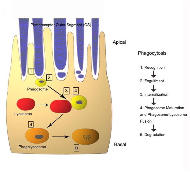
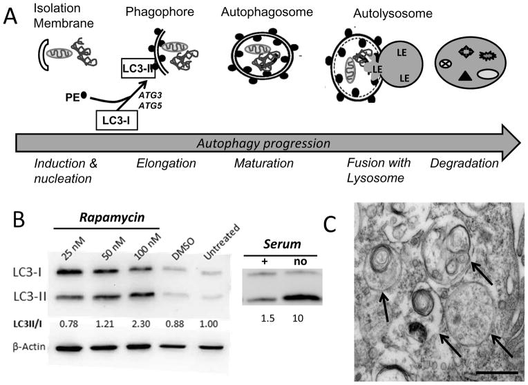
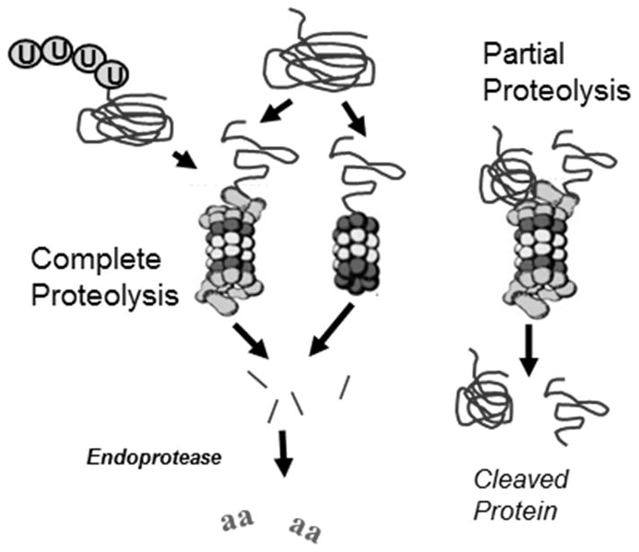
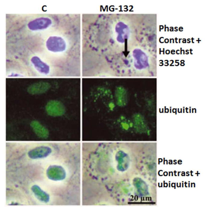
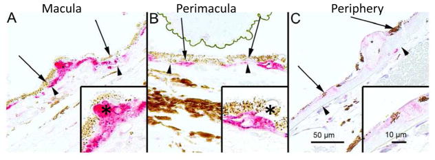
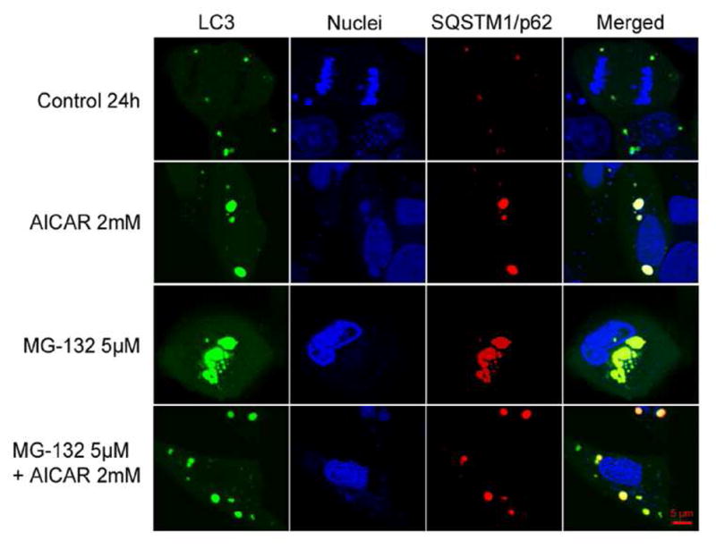
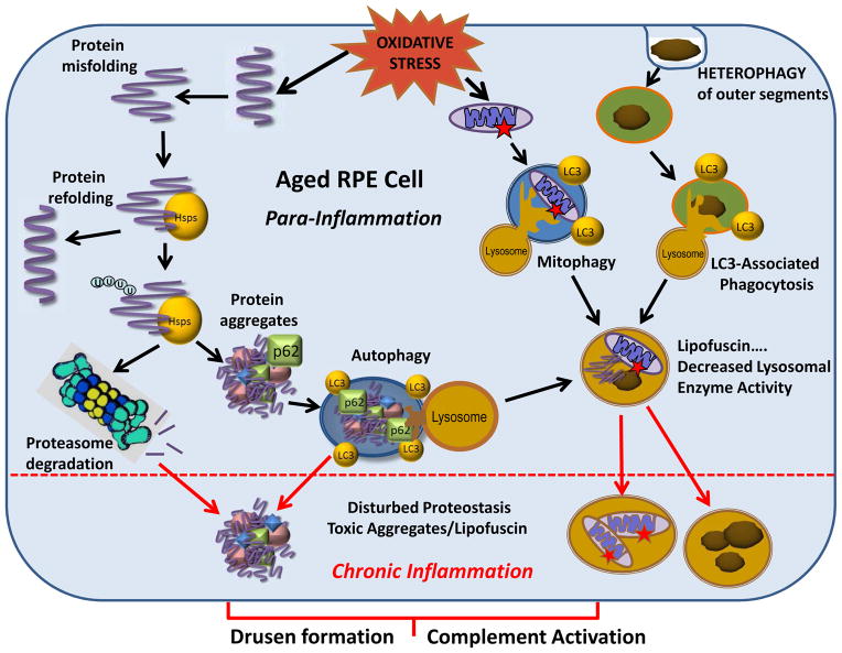
Maintenance of protein homeostasis, also referred to as "Proteostasis", integrates multiple pathways that regulate protein synthesis, folding, translocation, and degradation. Failure in proteostasis may be one of the underlying mechanisms responsible for the cascade of events leading to age-related macular degeneration (AMD). This review covers the major degradative pathways (ubiquitin-proteasome and lysosomal involvement in phagocytosis and autophagy) in the retinal pigment epithelium (RPE) and summarizes evidence of their involvement in AMD. Degradation of damaged and misfolded proteins via the proteasome occurs in coordination with heat shock proteins. Evidence of increased content of proteasome and heat shock proteins in retinas from human donors with AMD is consistent with increased oxidative stress and extensive protein damage with AMD. Phagocytosis and autophagy share key molecules in phagosome maturation as well as degradation of their cargo following fusion with lysosomes. Phagocytosis and degradation of photoreceptor outer segments ensures functional integrity of the neural retina. Autophagy rids the cell of toxic protein aggregates and defective mitochondria. Evidence suggesting a decline in autophagic flux includes the accumulation of autophagic substrates and damaged mitochondria in RPE from AMD donors. An age-related decrease in lysosomal enzymatic activity inhibits autophagic clearance of outer segments, mitochondria, and protein aggregates, thereby accelerating the accumulation of lipofuscin. This cumulative damage over a person's lifetime tips the balance in RPE from a state of para-inflammation, which strives to restore cell homeostasis, to the chronic inflammation associated with AMD.

Keywords: Age-related macular degeneration; Autophagy; Lysosome; Proteasome; Proteostasis; Retinal pigment epithelium.


Figures

Figure 1
Figure 1. Schematic depiction of phagocytosis in the RPE
The photoreceptor outer segment phagocytosis process includes outer segment recognition (1), engulfment (2), internalization (3), maturation and fusion with lysosomes (4) and final degradation within the phagolysosome (5). Please refer to the text for detailed information on the different stages of phagocytosis process.
Figure 2
Figure 2. Disrupted phagocytosis with Nuc1 depletion
(A, B) Transmission electron microscopy of RPE from 1-yr old wild type rats shows normal accumulation of lipofuscin-like particles (arrows) in the apical cytoplasm of RPE cells. (C) At higher magnification, RPE cells from 1 year old Nuc1 animals show abundant lipofuscin-like aggregates in the cytoplasm (arrows) and a large phagosome containing undigested outer segment discs (arrowhead). Scale bar = 500 nm. Reprinted with permission of J Cell Science.
Figure 3
Figure 3. Age-dependent abnormalities in the RPE cells of Cryba1 cKO mice
Transmission electron microscopy of 2-month old cKO RPE shows many vacuole-like structures with degenerated membrane-bound cellular organelles (arrows, center), undigested photoreceptor outer segments (arrow, right) and loss and truncation of basal infoldings (asterisks), as compared to Cryba1fl/fl (left). Scale bar= 500 nm. Reprinted with permission of Autophagy.
Fig. 4
Fig. 4
Time course of autophagy induction. (A) Steps in the autophagic degradation of cell contents include the induction of the isolation membrane followed by elongation of the double membrane to form the phagophore, which begins surrounding damaged organelles (eg., mitochondria) and protein aggregates. The lipidation of LC3-I by phosphatidylethanolamine (PE) to form LC3-II, which binds to the forming double membrane structure, is a crucial step in the maturation of the autophagosome. A commonly used indicator of autophagosome formation is the increase in LC3II. The autolysosome occurs when the autophagosome merges with a lysosome, after which lysosomal enzymes (LE) degrade the cargo. (B) Autophagic flux can be induced by inhibiting the mTOR pathway with rapamycin or under conditions of starvation (no serum). On Western blots, the lipidation by PE alters the migration of LC3 and permits quantification of both unlipidated (LC3-I) and lipidated (LC3-II) forms of the protein. Numbers below the figure are the ratio from densitometry of individual protein bands. Two examples of autophagy induction in primary cultures of human RPE show an increase in both total LC3 content and the lipidated form of the protein. At 24 hrs post-rapamycin treatment, hRPE show a dose-dependent increase in the LC3 II/I ratio. Serum deprivation for 2 hrs also dramatically increased the LC3 II/I ratio. Unpublished data. (C) The presence of the double membrane (arrows) verifies autophagosome formation in ARPE-19 cells 24 hrs after treatment with 50 nM bafilomycin. Scale bar =500 nm. Unpublished data.
Figure 5
Figure 5. Rab7 expression with p62 silencing and proteasome inhibition
Summary of densitometry from Western blots of Rab7 in homogenates of WT or SQSTM1/p62 silenced ARPE-19 cells after exposure to 1 μM MG-132 for 24 h. Results are expressed as means ± S.E.M. **p < 0.05 by Mann-Whitney test for the indicated comparisons. Unpublished data.
Figure 6
Figure 6. LAMP-2 expression under proteasome inhibition
Phase contrast microscopy and immunofluorescence staining of LAMP-2 in control cells, and cells exposed to 10 μM MG-132 for 24 hrs. Nuclei were stained with Hoechst 33258 dye (blue). Reprinted with permission of Journal of Cellular and Molecular Medicine.
Figure 7
Figure 7. Mitophagic elimination of damaged mitochondria
Steps for segregation and elimination of damaged mitochondria includes: (1) Mitochondrial damage (star) followed by fission isolates damaged mitochondria. (2) Pink1 accumulates on damaged mitochondria and recruits Parkin, which ubiquitinates mitofusion. (3) Ub-mitofusion is degraded by the proteasome. (4) Reduced mitofusion prevent fusion of damaged with healthy mitochondria. (5) Damaged mitochondria are targeted for mitophagy.
Figure 8
Figure 8. Immunostaining for SQSTM1/p62 in human AMD samples
The extent of cytoplasmic immunopositivity in the retinal pigment epithelial cells (RPE, shown by arrows) and in the drusen was evaluated microscopically (no staining or positive staining) by selecting 5 mm long areas of foveomacular (A), perimacular (B) and peripheral (C) regions. The drusen (shown by asterisks) were mostly SQSTM1/p62 negative. The nuclei of RPE cells were SQSTM1/p62 negative. (Original magnifications of x 200 and in insets x 400; Bruch’s membrane shown by arrow heads). Reprinted with permission of PlosOne.
Figure 9
Figure 9. Proteasome structure and regulatory proteins
The 20S core particle contains the standard (β1, β2, β5), immunoproteasome (LMP2, MECL-1, LMP7) catalytic subunits, or a mixture of both types of catalytic subunits. The 20S can bind to the regulatory complexes PA700 and/or PA28 to form the 26S, hybrid proteasome, or the immunoproteasome.
Figure 10
Figure 10. RPE Immunoproteasome induction by conditions of stress
Immunoproteasome content was upregulated in cultures of WT murine RPE following exposure to (A) chronic, low levels of hydrogen peroxide (0.5 mM), (B) TNFα (1 ng/ml) to mimic the inflammatory response, and (C) tunicamycin (5 μg/ml) to induce ER stress. Blots show antibody reaction to immunoproteasome subunits LMP2 and LMP7. Total proteasome content was estimated from reactions of alpha subunits (α6, α7), which are present in all proteasome subtypes. (A, B) Graph and numbers below blots indicate the densitometry of immune reactions relative to untreated controls. (C) Graph shows the dose-dependent response of RPE cell viability for WT and cells lacking the LMP7 and MECL-1 (L7M1) immunoproteasome subunits. Unpublished data.
Figure 11
Figure 11. Ubiquitin conjugation marks proteins for proteasome degradation
The coordinate action of the enzymes E1, E2, and E3 actively conjugate ubiquitin proteins to a lysine (K) residue on the protein substrate to mark it for proteasome degradation. An ubiquitin chain of at least 4 proteins is required for recognition by the regulatory complex of the 26S proteasome. Following binding of the Ub-protein, the regulatory complex deubiquitinates the substrate prior to protein unfolding and translocation into the core, where it is degraded to small peptides. The ubiquitin is recycled for another round of protein marking.
Figure 12
Figure 12. Signals for degradation by the proteasome
(A) PEST sequences are degrons that are buried within the proteins interior. With modification of the protein, such as phosphorylation, there is a change in protein structure that causes the cryptic sequence to become solvent exposed. The degron is recognized by a specific E3 ligase, which targets the protein for proteasome degradation. (B) Protein modification, such as oxidation, causes a change in protein structure that exposes hydrophobic residues that are normally buried within the protein. Exposure of hydrophobic patches targets this molecule for proteasome degradation through a mechanism that is still unclear.
Figure 13
Figure 13. Proteolytic degradation of protein substrates by the proteasome
Proteins containing a degradation signal (eg., ubiquitin modification or exposed hydrophobic amino acids) are targeted to the proteasome. Complete proteolysis results in the release of multiple peptides of varying size. These peptides are further degraded to amino acids by cellular endopeptidases. Partial proteolysis occurs when an unstructured region of a protein gains access to catalytic sites within the 20S core. Enzymatic cleavage cuts the protein so that two partial pieces of the protein are released.
Figure 14
Figure 14. Ubiquitinated protein conjugates accumulate under proteasome inhibition
Phase contrast microscopy and immunofluorescence analysis for ubiquitin (green) in control cells, and cells exposed to 10 μM MG-132 proteasome inhibition for 24 hrs. Nuclei were stained with Hoechst 33258 dye (blue). Arrows point to perinuclear protein aggregates. Reprinted with permission of Journal of Cellular and Molecular Medicine.
Figure 15
Figure 15. Immunostaining for ubiquitin in human AMD samples
The ubiquitin immune reaction in the retinal pigment epithelial cells (RPE, shown by arrows) and in the drusen (asterisks) was evaluated microscopically by selecting foveomacular (A), perimacular (B) and peripheral (C) regions. Most of the drusen were strongly ubiquitin-positive (asterisks). Bruch’s membrane is shown by arrow heads. Original magnification was 200x and in insets 400x. Reprinted with permission of PlosOne.
Figure 16
Figure 16. Co-localization of LC3 and SQSTM1/p62
Confocal images show staining for LC3 (pDendra2-hLC3, green) and SQSTM1/p62 (pDsRed2-hp62, red), and nuclei (Hoechst 33258 dye, blue) in ARPE-19 cells. An orange/yellow signal in the merged image indicates co-localization of LC3 and SQSTM1/p62 in untreated control ARPE-19 cells and cells exposed to 2 mM AICAR or/and 5 μM MG-132 for 24 h. Reprinted with permission of PlosOne.
Figure 17
Figure 17. Hsp70 expression under proteasome inhibition
(A) Phase contrast microscopy and immunofluorescence analysis for Hsp70 (green) in control cells, and cells exposed to 10 μM MG-132 proteasome inhibition for 24 hrs. Nuclei were stained with Hoechst 33258 dye (blue). Arrows point to perinuclear protein aggregates. (B) Western blotting analysis (10 μg protein/lane) of the Hsp70 content in isolated lysosome fractions from the control cells (lanes for C) or cells exposed to 10 μM MG-132 for 24hrs (lane 0) or cells exposed to 10 μM MG-132 for 24 and then allowed to recover for up to 48 hrs (lanes 24 and 48). Lysate from heat shocked cells (H/S) are the positive control. Reprinted with permission of Journal of Cellular and Molecular Medicine.
Figure 18
Figure 18. Dysregulated proteostasis in aged RPE cells evokes AMD pathology
RPE cells are constantly exposed to oxidative stress. One consequence is that damage to proteins causes their unfolding. Heat-shock proteins (Hsps) attempt to refold damaged proteins, but if not successful, the misfolded proteins are ubiquitinated (U) and targeted for proteasomal clearance. If proteasome activity is decreased, proteins aggregate and are degraded via autophagy with the assistance of p62. Autophagy is also used to digest damaged mitochondria (mitophagy). Photoreceptor outer segments that are phagocytosed by the RPE (heterophagy) are degraded in the lysosomes in a recently described process of LC3-Associated Phagocytosis. In aged RPE cells, lipofuscin accumulates in lysosomes as a result of the coincident decline of lysosomal enzyme activity. The impaired lysosomal enzyme activity inhibits autophagic flux and non-digested mitochondria, protein aggregates, and lipofuscin accumulate. Thus, in aged RPE cells, disturbed proteostasis and accumulated toxic compounds trigger the progression from para-inflammation to chronic inflammation and evoke the AMD-associated formation of extracellular drusen formation and complement activation.

Similar articles

Cited by

References

    1. Ambati J, Atkinson JP, Gelfand BD. Immunology of age-related macular degeneration. Nat Rev Immunol. 2013;13:438–451. - PMC - PubMed
    1. Anderson DH, Radeke MJ, Gallo NB, Chapin EA, Johnson PT, Curletti CR, Hancox LS, Hu J, Ebright JN, Malek G, Hauser MA, Rickman CB, Bok D, Hageman GS, Johnson LV. The pivotal role of the complement system in aging and age-related macular degeneration: hypothesis re-visited. Prog Retin Eye Res. 2010;29:95–112. - PMC - PubMed
    1. Agarwal AK, Xing C, DeMartino GN, Mizrachi D, Hernandez MD, Sousa AB, Martinez de Villarreal L, dos Santos HH, Garg A. PSMB8 encoding the β5i proteasome subunit is mutated in joint contractures, muscle atrophy, microcytic anemia, and panniculitis-induced lipodystrophy syndrome. Am J Hum Genet. 2010;87:866–872. - PMC - PubMed
    1. Arima K, Kinoshita A, Mishima H, Kanazawa N, Kaneko T, Mizushima T, et al. Proteasome assembly defect due to a proteasome subunit beta type 8 (PSMB8) mutation causes the autoinflammatory disorder, Nakajo-Nishimura syndrome. Proc Natl Acad Sci U S A. 2011;108:14914–14919. - PMC - PubMed
    1. Ban T, Heymann JAW, Song Z, Hinshaw JE, Chan DC. OPA1 disease alleles causing dominant optic atrophy have defects in cardiolipin-stimulated GTP hydrolysis and membrane tabulation. Hum Mol Genet. 2010;19:2113–2122. - PMC - PubMed

Publication types

Substances