. 2015 Nov 6;427(22):3587-3597.
doi: 10.1016/j.jmb.2015.09.001. Epub 2015 Sep 5.

Effects of Linker Length and Transient Secondary Structure Elements in the Intrinsically Disordered Notch RAM Region on Notch Signaling

Affiliations

Effects of Linker Length and Transient Secondary Structure Elements in the Intrinsically Disordered Notch RAM Region on Notch Signaling

Kathryn P Sherry et al. J Mol Biol. .

Abstract

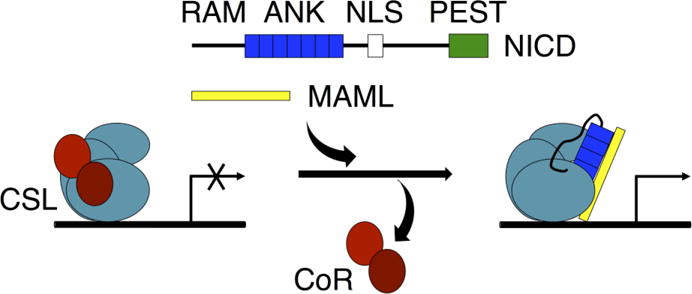
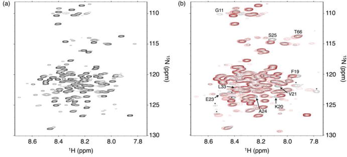
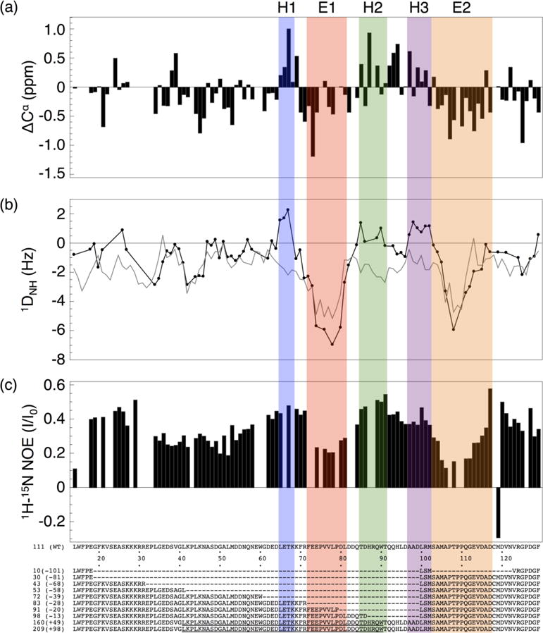
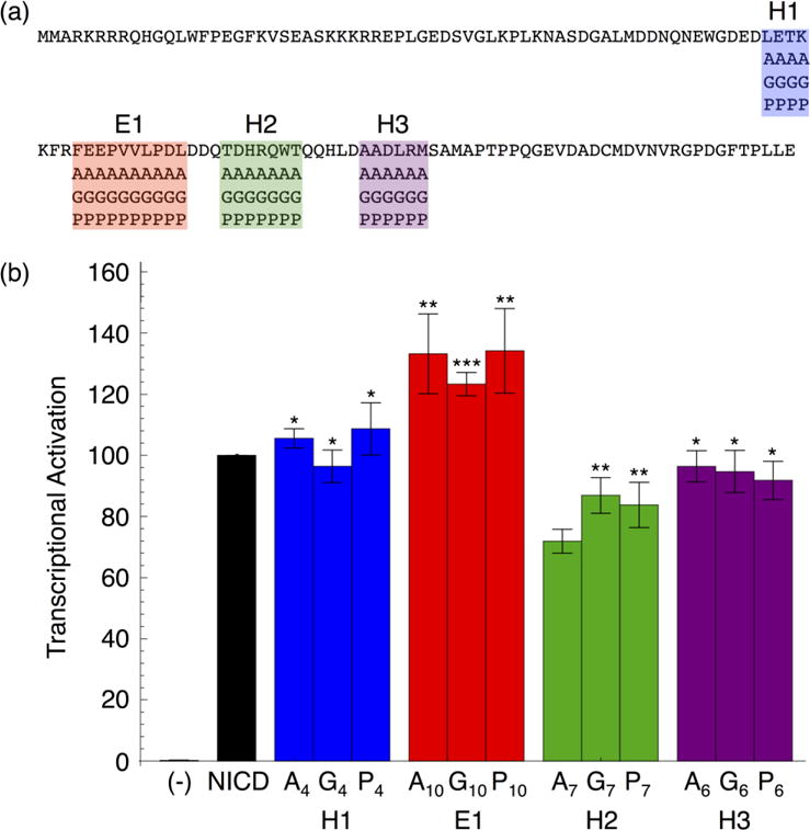
Formation of the bivalent interaction between the Notch intracellular domain (NICD) and the transcription factor CBF-1/RBP-j, Su(H), Lag-1 (CSL) is a key event in Notch signaling because it switches Notch-responsive genes from a repressed state to an activated state. Interaction of the intrinsically disordered RBP-j-associated molecule (RAM) region of NICD with CSL is thought to both disrupt binding of corepressor proteins to CSL and anchor NICD to CSL, promoting interaction of the ankyrin domain of NICD with CSL through an effective concentration mechanism. To quantify the role of disorder in the RAM linker region on the effective concentration enhancement of Notch transcriptional activation, we measured the effects of linker length variation on activation. The resulting activation profile has general features of a worm-like chain model for effective concentration. However, deviations from the model for short sequence deletions suggest that RAM contains sequence-specific structural elements that may be important for activation. Structural characterization of the RAM linker with sedimentation velocity analytical ultracentrifugation and NMR spectroscopy reveals that the linker is compact and contains three transient helices and two extended and dynamic regions. To test if these secondary structure elements are important for activation, we made sequence substitutions to change the secondary structure propensities of these elements and measured transcriptional activation of the resulting variants. Substitutions to two of these nonrandom elements (helix 2, extended region 1) have effects on activation, but these effects do not depend on the nature of the substituting residues. Thus, the primary sequences of these elements, but not their secondary structures, are influencing signaling.

Keywords: NMR spectroscopy; analytical ultracentrifugation; intrinsically disordered proteins; protein–protein interactions; reporter gene assays.


Figures

Figure 1
Figure 1. Schematic of Notch signaling
NICD (containing RAM, ANK, NLS, and PEST regions) enters the nucleus after it is cleaved from the Notch receptor upon receptor activation. The RAM region and ANK domain of NICD bind to CSL at the promoter region of Notch target genes. Binding of NICD to CSL disrupts binding of co-repressor (CoR) proteins and promotes binding of the co-activator protein MAML.
Figure 2
Figure 2. Transcription activities of RAM linker sequence deletions and duplications
(a) Schematic of NICD and constructs with sequence deletions and duplications in the RAM region. Each construct is labeled with the length, in residues, of the RAM linker region and the number of residues deleted or inserted (in parentheses). The duplicated sequence (black arrow) was replicated once (+49) or twice (+98) (gray arrows). (b) NICD-mediated transcriptional activation (black) and effective concentration (gray) of constructs with different RAM linker lengths. Effective concentrations were calculated from the worm-like chain model.
Figure 3
Figure 3. 15N-1H HSQC spectra of uniformly labeled 15N RAM
(a) Spectrum of 15N RAM at 800 MHz. Most resonances are reasonably well dispersed for an IDP, facilitating assignment with standard triple resonance experiments. (b) Overlay of 15N RAM spectra with (red) and without (black) a 1.4:1 molar excess of CSL at 600 MHz. Resonances that move upon RAM:CSL binding are labeled and correspond to residues at the RAM N-terminus. Unassigned resonances that move are labeled with an asterisk. Conditions: 25 mM sodium phosphate pH 6.5, 50 mM sodium chloride, 0.1 mM TCEP, and 1 mM EDTA at 20 °C.
Figure 4
Figure 4. Local structural features of RAM determined by NMR spectroscopy
(a) Cα Chemical shift deviations from random coil values. (b) 15N-1H Amide residual dipolar coupling values (black circles), and values predicted by Flexible-Meccano (gray). (c) 1H-15N Heteronuclear NOE values. Based on these three parameters, we identify three transiently helical segments H1-H3 (blue, green and purple), and two extended and dynamic segments E1 and E2 (green and orange). The first twelve and last five residues of RAM are not shown because of missing assignments.
Figure 5
Figure 5. Transcription activities of block sequence substitutions in regions of RAM containing residual secondary structure
(a) RAM sequence with block substitution boundaries. Each RAM variant contains one helical or extended region substituted with alanine, glycine, or proline residues. (b) Transcriptional activation of RAM sequence substitutions in NICD. Substitutions in the E1 and H2 regions change transcriptional activation, but this change does not depend on secondary structure type. Error bars show the standard error of the mean and statistical significance compared to NICD is labeled as ***P < 0.001, **P < 0.01, and *P < 0.05.
Figure 6
Figure 6. Hydrodynamic radii (RH) of RAM determined by SV-AUC at various concentrations of NaCl
The solid line is a linear fit to the data. The dashed lines are RH values calculated from scaling relationships for folded and unfolded proteins and for intrinsically disordered proteins of the same molecular weight as RAM at physiological salt concentration.

Similar articles

Cited by

References

    1. Artavanis-Tsakonas S, Rand MD, Lake RJ. Notch signaling: cell fate control and signal integration in development. Science. 1999;284:770–6. - PubMed
    1. Kopan R, Ilagan MXG. The canonical Notch signaling pathway: unfolding the activation mechanism. Cell. 2009;137:216–33. - PMC - PubMed
    1. Gridley T. Notch signaling and inherited disease syndromes. Hum Mol Genet. 2003;12:9R–13. - PubMed
    1. Louvi A, Arboleda-Velasquez J, Artavanis-Tsakonas S. CADASIL: a critical look at a Notch disease. Dev Neurosci. 2006;28:5–12. - PubMed
    1. Weng AP, Ferrando AA, Lee W, Morris JP, Silverman LB, Sanchez-Irizarry C, Blacklow SC, Look AT, Aster JC. Activating mutations of NOTCH1 in human T cell acute lymphoblastic leukemia. Science. 2004;306:269–71. - PubMed

Publication types

MeSH terms

Substances

LinkOut - more resources