Comparative Study
. 2016 May;83(5):880-888.e2.
doi: 10.1016/j.gie.2015.08.050. Epub 2015 Sep 3.

Comparative diagnostic performance of volumetric laser endomicroscopy and confocal laser endomicroscopy in the detection of dysplasia associated with Barrett's esophagus

Affiliations
Comparative Study

Comparative diagnostic performance of volumetric laser endomicroscopy and confocal laser endomicroscopy in the detection of dysplasia associated with Barrett's esophagus

Cadman L Leggett et al. Gastrointest Endosc. 2016 May.

Abstract

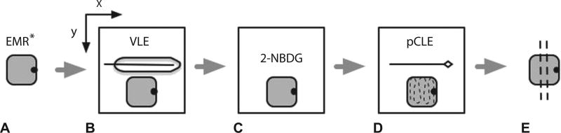
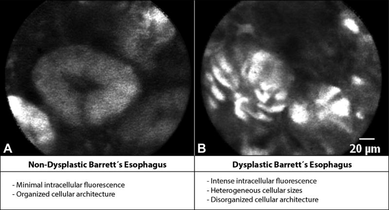
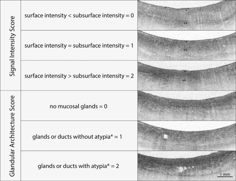
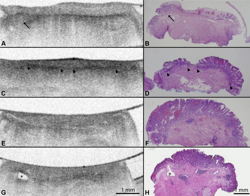
Background and aims: Probe-based confocal laser endomicroscopy (pCLE) and volumetric laser endomicroscopy (VLE) (also known as frequency domain optical coherence tomography) are advanced endoscopic imaging modalities that may be useful in the diagnosis of dysplasia associated with Barrett's esophagus (BE). We performed pCLE examination in ex-vivo EMR specimens and compared the diagnostic performance of using the current VLE scoring index (previously established as OCT-SI) and a novel VLE diagnostic algorithm (VLE-DA) for the detection of dysplasia.

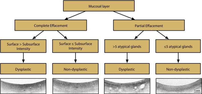
Methods: A total of 27 patients with BE enrolled in a surveillance program at a tertiary-care center underwent 50 clinically indicated EMRs that were imaged with VLE and pCLE and classified into neoplastic (N = 34; high-grade dysplasia, intramucosal adenocarcinoma) and nonneoplastic (N = 16; low-grade dysplasia, nondysplastic BE), based on histology. Image datasets (VLE, N = 50; pCLE, N = 50) were rated by 3 gastroenterologists trained in the established diagnostic criteria for each imaging modality as well as a new diagnostic algorithm for VLE derived from a training set that demonstrated association of specific VLE features with neoplasia. Sensitivity, specificity, and diagnostic accuracy were assessed for each imaging modality and diagnostic criteria.

Results: The sensitivity, specificity, and diagnostic accuracy of pCLE for detection of BE dysplasia was 76% (95% confidence interval [CI], 59-88), 79% (95% CI, 53-92), and 77% (95% CI, 72-82), respectively. The optimal diagnostic performance of OCT-SI showed a sensitivity of 70% (95% CI, 52-84), specificity of 60% (95% CI, 36-79), and diagnostic accuracy of 67%; (95% CI, 58-78). The use of the novel VLE-DA showed a sensitivity of 86% (95% CI, 69-96), specificity of 88% (95% CI, 60-99), and diagnostic accuracy of 87% (95% CI, 86-88). The diagnostic accuracy of using the new VLE-DA criteria was significantly superior to the current OCT-SI (P < .01).

Conclusion: The use of a new VLE-DA showed enhanced diagnostic performance for detecting BE dysplasia ex vivo compared with the current OCT-SI. Further validation of this algorithm in vivo is warranted.


Figures

Figure 1
Figure 1
Schematic representation of steps involved in imaging acquisition. A, EMR* specimen is marked for orientation. EMR specimen is placed on a holder attached to a translational mechanical stage that allows for movement in the x and y planes. B, EMR specimen is imaged with volumetric laser endomicroscopy. C, EMR specimen is incubated in 0.5 µM 2-[N-(7-nitrobenz-2-oxa-1,3-diaxol-4-yl)amino]-2-deoxyglucose for 20 minutes. D, EMR specimen is imaged with probe confocal laser endomicroscopy in video format across the entire surface area of the specimen in a grid scanning pattern. E, EMR specimen is placed in formaldehyde and submitted for histologic sectioning. EMR and imaging probes not to scale. VLE, volumetric laser endomicroscopy; 2-NBDG, 2-[N-(7-nitrobenz-2-oxa-1,3-diaxol-4-yl)amino]-2-deoxyglucose; pCLE, probe confocal laser endomicroscopy.
Figure 2
Figure 2
Probe confocal laser endomicroscopy (pCLE) fluorescence criteria. A, pCLE images of nondysplastic Barrett’s esophagus (BE) showing minimal intracellular fluorescence and organized cellular architecture. B, Dysplastic BE showing intense intracellular fluorescence with heterogeneous cellular sizes and disorganized cellular architecture based on the pCLE fluorescence criteria.
Figure 3
Figure 3
The volumetric laser endomicroscopy scoring index (OCT-SI) is used to diagnose Barrett’s esophagus (BE)-associated dysplasia. The scoring index consists of 2 independent criteria (surface-to-subsurface signal intensity and glandular architecture) that are added to calculate a dysplasia score. A dysplasia score of ≥2 is associated with a sensitivity of 83% and a specificity of 75% for the diagnosis of neoplasia in BE by using optical coherence tomography. The volumetric laser endomicroscopy surface signal intensity is represented by + and the subsurface intensity by ++. Glandular atypia is defined by the presence of irregular and/or dilated glands.
Figure 4
Figure 4
A, Volumetric laser endomicroscopy (VLE) imaging of EMR specimens with confirmed B, histopathology showing layered mucosal architecture of squamous epithelium (H&E, orig. mag. × 15). Black arrow shows a small focus of subsquamous Barrett’s esophagus (BE). C, D, VLE images of histopathology confirmed nondysplastic BE showing loss of layered mucosal architecture and partial effacement of the mucosal layer (>2-mm layer). The partial effacement of the VLE mucosal layer shows less distinction between mucosa and submucosa compared with squamous epithelium (arrowheads). E, VLE images of BE with high-grade dysplasia confirmed by F, histology showing loss of layered mucosal architecture and complete effacement of the mucosal layer (<2-mm layer). G, VLE images of histologically confirmed high-grade dysplasia showing an atypical glandular structure that represents a dilated submucosal duct (as noted by the asterisk).
Figure 5
Figure 5
Volumetric laser endomicroscopy diagnostic algorithm (VLE-DA). Interpretation of the VLE-DA is performed over a longitudinal distance of 1 cm of Barrett’s esophagus (BE). Partial effacement of the mucosal layer is defined by a mucosal layer ≥2 mm in transverse cross-section present in ≥50% of the scan. Complete effacement of the mucosal layer is defined by the presence of a mucosal layer over <50% of the scan. A rating of image surface-to-subsurface intensity corresponds to the most prevalent ratio (surface > subsurface intensity vs surface < subsurface intensity) present in ≥50% of the scan. The VLE surface signal intensity is represented by + and subsurface intensity by ++.
Figure 6
Figure 6
A, Graph showing diagnostic accuracy of point confocal laser endomicroscopy compared with volumetric laser endomicroscopy (VLE) by using the VLE scoring index (OCT-SI) at a diagnostic threshold of ≥3 and the diagnostic algorithm. An OCT-SI threshold of ≥3 showed improved diagnostic characteristics compared with other thresholds on post hoc analysis. B, Table showing sensitivity and specificity of pCLE, OCT-SI, and VLE-DA. pCLE, probe confocal laser endomicroscopy; OCT-SI, VLE scoring index; VLE-DA, volumetric laser endomicroscopy diagnostic algorithm; CI, confidence interval; PPV, positive predictive value; NPV, negative predictive value.

References

    1. Pohl H, Welch HG. The role of overdiagnosis and reclassification in the marked increase of esophageal adenocarcinoma incidence. J Natl Cancer Inst. 2005;97:142–6. - PubMed
    1. Qumseya BJ, Wang H, Badie N, et al. Advanced imaging technologies increase detection of dysplasia and neoplasia in patients with Barrett’s esophagus: a meta-analysis and systematic review. Clin Gastroenterol Hepatol. 2013;11:1562–1570. e1–2. - PMC - PubMed
    1. Sharma P, Meining AR, Coron E, et al. Real-time increased detection of neoplastic tissue in Barrett’s esophagus with probe-based confocal laser endomicroscopy: final results of an international multicenter, prospective, randomized, controlled trial. Gastrointest Endosc. 2011;74:465–72. - PMC - PubMed
    1. Sauk J, Coron E, Kava L, et al. Interobserver agreement for the detection of Barrett’s esophagus with optical frequency domain imaging. Dig Dis Sci. 2013;58:2261–5. - PMC - PubMed
    1. Evans JA, Poneros JM, Bouma BE, et al. Optical coherence tomography to identify intramucosal carcinoma and high-grade dysplasia in Barrett’s esophagus. Clin Gastroenterol Hepatol. 2006;4:38–43. - PMC - PubMed

Publication types