. 2015 Sep 7:14:83.
doi: 10.1186/s12938-015-0076-7.

A new system for quantitative evaluation of infant gaze capabilities in a wide visual field

Affiliations

A new system for quantitative evaluation of infant gaze capabilities in a wide visual field

Andrea Pratesi et al. Biomed Eng Online. .

Abstract

Background: The visual assessment of infants poses specific challenges: many techniques that are used on adults are based on the patient's response, and are not suitable for infants. Significant advances in the eye-tracking have made this assessment of infant visual capabilities easier, however, eye-tracking still requires the subject's collaboration, in most cases and thus limiting the application in infant research. Moreover, there is a lack of transferability to clinical practice, and thus it emerges the need for a new tool to measure the paradigms and explore the most common visual competences in a wide visual field. This work presents the design, development and preliminary testing of a new system for measuring infant's gaze in the wide visual field called CareToy C: CareToy for Clinics.

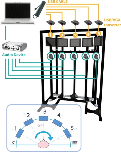
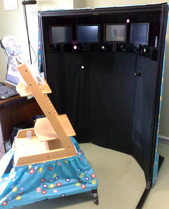
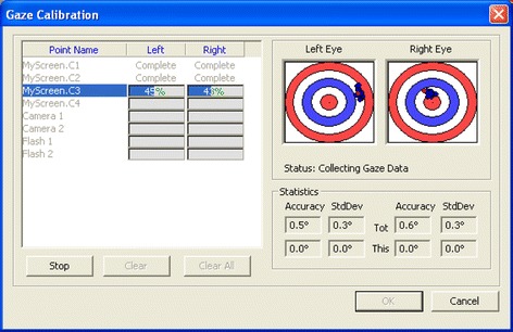
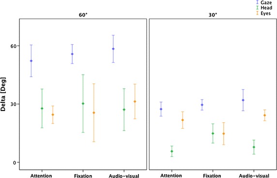
Methods: The system is based on a commercial eye tracker (SmartEye) with six cameras running at 60 Hz, suitable for measuring an infant's gaze. In order to stimulate the infant visually and audibly, a mechanical structure has been designed to support five speakers and five screens at a specific distance (60 cm) and angle: one in the centre, two on the right-hand side and two on the left (at 30° and 60° respectively). Different tasks have been designed in order to evaluate the system capability to assess the infant's gaze movements during different conditions (such as gap, overlap or audio-visual paradigms). Nine healthy infants aged 4-10 months were assessed as they performed the visual tasks at random.

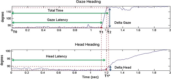
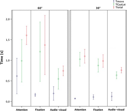
Results: We developed a system able to measure infant's gaze in a wide visual field covering a total visual range of ±60° from the centre with an intermediate evaluation at ±30°. Moreover, the same system, thanks to different integrated software, was able to provide different visual paradigms (as gap, overlap and audio-visual) assessing and comparing different visual and multisensory sub-competencies. The proposed system endowed the integration of a commercial eye-tracker into a purposive setup in a smart and innovative way.

Conclusions: The proposed system is suitable for measuring and evaluating infant's gaze capabilities in a wide visual field, in order to provide quantitative data that can enrich the clinical assessment.


Figures

Fig. 1
Fig. 1
Schematic system overview. The mechanical custom structure represents the support for the five screens, the five speakers, the six SmartEye cameras running at 60 Hz and two IR-diodes used for illuminating the face of the subject in order to minimize the effect of varying environmental lighting conditions and for using the reflections of these IR flashes on the cornea (“glints”) to find the centre of the eyes. The stimuli management has been obtained using a laptop combined with the audio–video external devices. In the lower part of this overview, it is possible to observe the gaze heading frame of reference
Fig. 2
Fig. 2
SmartEye Graphical User Interface (GUI). In the upper bar it is possible see the pink vectors that represent the infant’s gaze vector; in the lower window on the left, there is the 3D representation of the external 3D setup with the visualisation of gaze intersection on a object modelled in the 3D world (i.e. the gaze vector intersects screen n.3) and on the right there are the typical gaze heading and head heading signal profiles
Fig. 3
Fig. 3
The final version of the system
Fig. 4
Fig. 4
Gaze heading and head heading signal profiles. An example of gaze heading and head heading time response with relative Total Time (TT), Gaze Latency (GL) and Head Latency (HL) parameters
Fig. 5
Fig. 5
Gaze calibration procedure. The red dot shows where the current un-calibrated gaze intersects a plane, orthogonal to the current world point and a vector pointing towards the centre of the eye. The blue dots represent all saved samples, whereas the green dots show the samples once the calibration algorithm has been run on them. Ideally the green dots should be in the middle of both the target. One circle in the target corresponds to ±2° of accuracy. We carefully checked that the blue dots were close together without outliers, and any outliers that were found were cleared and new samples were added again. We manually repeated this operation until the noise became smaller
Fig. 6
Fig. 6
Gaze heading signal, velocity and acceleration profile
Fig. 7
Fig. 7
Examples of attention task: a results of SmartEye analysis of intersection between the gaze with the screens. During the transition from screen #3 to #4, the gaze passes through the space between the two screens thus it does not intersect one of the AOI and the system returns zero value, b gaze heading during the transition from screen #3 to screen #4, c head heading during the transition from screen #3 to screen #4, d results of SmartEye analysis of intersection between the gaze with the peripheral screens. In this case the system returns zero value when the gaze is between screen 3 and 4 and between 4 and 5, e gaze heading during the transition from screen #3 to screen #5, f head heading during the transition from screen #3 to screen #5
Fig. 8
Fig. 8
Time parameters results
Fig. 9
Fig. 9
Delta parameters results

References

    1. Dubowitz LM, Dubowitz V, Morante A. Visual function in the newborn: a study of preterm and full-term infants. Brain Dev. 1980;2:15–29. doi: 10.1016/S0387-7604(80)80004-0. - DOI - PubMed
    1. Madan A, Jan JE, Good WV. Visual development in preterm infants. Dev Med Child Neurol. 2005;47:276–280. doi: 10.1017/S0012162205000514. - DOI - PubMed
    1. Morante A, Dubowitz LM, Leven M, Dubowitz V. The development of visual function in normal and neurologically abnormal preterm and fullterm infants. Dev Med Child Neurol. 1982;24:771–784. doi: 10.1111/j.1469-8749.1982.tb13698.x. - DOI - PubMed
    1. Gredebäck G, Johnson S, von Hofsten C. Eye tracking in infancy research. Dev Neuropsychol. 2010;35:1–19. doi: 10.1080/87565640903325758. - DOI - PubMed
    1. Corbetta D, Guan Y, Williams JL. Infant eye-tracking in the context of goal-directed actions. Infancy. 2012;17:102–125. doi: 10.1111/j.1532-7078.2011.00093.x. - DOI - PMC - PubMed

Publication types

LinkOut - more resources