. 2015 Sep 8:5:13774.
doi: 10.1038/srep13774.

Adenylate Cyclase Toxin promotes bacterial internalisation into non phagocytic cells

Affiliations

Adenylate Cyclase Toxin promotes bacterial internalisation into non phagocytic cells

César Martín et al. Sci Rep. .

Abstract

Bordetella pertussis causes whooping cough, a respiratory infectious disease that is the fifth largest cause of vaccine-preventable death in infants. Though historically considered an extracellular pathogen, this bacterium has been detected both in vitro and in vivo inside phagocytic and non-phagocytic cells. However the precise mechanism used by B. pertussis for cell entry, or the putative bacterial factors involved, are not fully elucidated. Here we find that adenylate cyclase toxin (ACT), one of the important toxins of B. pertussis, is sufficient to promote bacterial internalisation into non-phagocytic cells. After characterization of the entry route we show that uptake of "toxin-coated bacteria" proceeds via a clathrin-independent, caveolae-dependent entry pathway, allowing the internalised bacteria to survive within the cells. Intracellular bacteria were found inside non-acidic endosomes with high sphingomyelin and cholesterol content, or "free" in the cytosol of the invaded cells, suggesting that the ACT-induced bacterial uptake may not proceed through formation of late endolysosomes. Activation of Tyr kinases and toxin-induced Ca(2+)-influx are essential for the entry process. We hypothesize that B. pertussis might use ACT to activate the endocytic machinery of non-phagocytic cells and gain entry into these cells, in this way evading the host immune system.


Figures
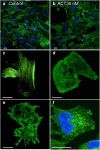
Figure 1
Figure 1. Reorganization of the cell cytoskeleton induced by ACT, or by B. pertussis.
Exposure of CHO-K1 cells to purified ACT or to B. pertussis visibly affected cellular actin microfilament organization. CHO-K1 cells were treated with purified ACT or with B. pertussis, fixed, permeabilized and stained with Alexa Fluor® 488 phalloidin to visualize actin cytoskeleton and with Hoechst to visualize DNA as described in Experimental procedures. (a) Control. Untreated CHO-K1 cells at low magnification; (b) CHO-K1 cells incubated with 2 μg/mL ACT, low magnification view; (c) More detailed view of control, untreated CHO-K1 cells shown at higher magnification; (d) Detailed view of CHO-K1 cells incubated with 2 μg/mL ACT and shown at higher magnification; (e) Detailed view of CHO-K1 cells incubated with 5 μg/mL ACT (f) Detailed view of CHO-K1 cells incubated with B. pertussis (m.o.i 100), shown at higher magnification in order to better visualize the bacteria stained by Hoechst. Representative confocal images from three independent experiments are shown. Scale bars, 20 μm. A minimum of 30 cells were analyzed in each individual experiment.
Figure 2
Figure 2. “ACT-coated bacteria” or virulent B. pertussis are taken up by non-phagocytic cells.
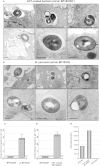
Incubation of CHO-K1 cells with “ACT-coated bacteria” (a) or with virulent, parental B. pertussis (b), results in bacterial internalisation, as visualized by electron microscopy. A detailed analysis of electron micrographs taken from infected CHO-K1 cells shows the different steps of the invasion process: first the bacteria were attached onto the cell membrane (upper left panel, 2a,b), then internalized, with some of the bacteria appearing inside vesicles, surrounded by a membrane (upper middle and upper right panels, 2a,b), and some of them appearing “free” in the cytosol (lower panels, 2a,b). Representative images of three independent experiments are shown. Scale bars, 0.5 μm. The bacterial invasion assay and electron microscopy analysis were performed as described in Experimental procedures. Panels (c,d) show respectively, the extent of bacterial internalisation quantified from cells treated with virulent parental B. pertussis (wt BP18323) relative to non-virulent strain (BP18323H-) and expressed as number of intracellular bacteria per cell, or from cells incubated with “ACT-coated bacteria” (BP18323H-+ACT) relative to non-virulent non-coated strain (BP18323H-), and expressed as number of intracellular bacteria per cell in both cases. Panel (e) shows the total number of internalized bacteria quantified in the three different cases after 2h of invasion.
Figure 3
Figure 3. Low magnification confocal images of CHO-K1 cells incubated with non-virulent B. pertussis (strain BP18323H-) or “ACT-coated bacteria”.
CHO-K1 cells were incubated with non-virulent B. pertussis (strain BP18323H-) (a) or with “ACT-coated bacteria” (b) and invasion assay and confocal microscopy analysis were performed as described in Experimental procedures. As observed in the images the non-virulent B.p. strain, which does not express nor ACT nor adhesins, was hardly found in the cell interior (a). In contrast, the non-virulent bacteria that are coated with ACT can be found in the interior of CHO-K1 cells. Cell membranes were stained with the fluorescent probe DiI (red) and DNA with Hoechst (blue). Representative images of three independent experiments are shown. A minimum of 30 cells were analyzed in each individual experiment. Scale bars, 20 μm.
Figure 4
Figure 4. Quantification of the total number of internalized bacteria and viability of the bacteria-containing cells.
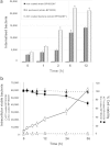
CHO-K1 cells were incubated with control non-virulent B. pertussis (BP18323H-), with virulent B. pertussis (strain BP18323) or with “ACT-coated bacteria”, and invasion was followed as described in Experimental procedures. The number of internalized bacteria at different times of invasion (0.5–12 h) was quantified as described in Experimental procedures. Time =0 h represents the invasion occurring after 2 h cell incubation with bacteria, washing and killing of extracellular bacteria by gentamicin (a). Cell viability of CHO-K1 cells and number of internalized bacteria were determined for each time-point (0.5–36 h) as described in Experimental procedures (b). Internalisation of “ACT-coated bacteria” only induced a slight to moderate decrease in the cell viability of the infected CHO-K1 cells, and most of the internalized bacteria could grow after infection. Data shown are the mean ± SD of at least three independent experiments performed in triplicate, with *p < 0.01.
Figure 5
Figure 5. Characterization of the invasion pathway of “ACT-coated bacteria”.
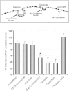
Several inhibitors of the different possible entry routes were analyzed and it was concluded that “ACT-coated B. pertussis” exploits a clathrin-independent, cholesterol-dependent (caveolae-dependent) entry pathway to invade CHO-K1 cells, and that it requires the activation of tyrosine kinases. CHO-K1 cells were pre-incubated with chemical inhibitors (5 μg/mL chlorpromazine, 450 mM sucrose, 10 mM methyl-β-cyclodextrin, 0.25 μg/mL nystatin, 100 μM genistein and 1 μM okadaic acid) for 30 min at 37 °C, then cell invasion was assayed as described in Experimental procedures. Chlorpromazine, sucrose and methyl-β-cyclodextrin were dissolved in DMEM, nystatin, genistein and okadaic acid, were dissolved in DMSO. Controls showing that vehicle has no effect on internalisation are shown in Fig. S5. The data were normalized to the control sample (no addition) and expressed as per cent of control entry. Data shown are the mean ± SD of at least three independent experiments performed in quintuplicate, with *p < 0.01.
Figure 6
Figure 6. Characterization of bacteria-containing endosomes.
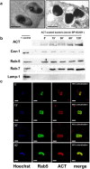
Ultrathin cuts of infected cells (a) or endosome membranes isolated from the infected cells at different times of incubation (5–120 min) and submitted to SDS-PAGE electrophoresis (b), or purified endosomes isolated and purified from the infected cells (c), were respectively analysed by electron microscopy (a), by Western blot (b) or by confocal microscopy (c). Common markers of the different endocytic stages were used for the respective analysis: gold-labelled anti-Rab-5 Ab for the electron microscopy (a), or anti-Rab-5 Ab, as early endosome marker; anti- Rab7 Ab as late endosomal marker, anti-LAMP-1 Ab as lysosomal marker, and anti-Cav-1 Ab as caveolae marker, in the Western blot assay (b). For confocal analysis DNA was stained with Hoechst, Rab-5 was stained with a FITC-labelled secondary Ab, and ACT was stained with a Texas Red®-labelled secondary Ab. Quantification of signal co-localization is given (c). From the combination of all the data it was concluded that “ACT-coated B. pertussis” resides in Cav-1 positive, Rab-5 and Rab-7 positive and LAMP-1 negative non-acidic compartments in the infected CHO-K1 cells. Representative images of three independent experiments are shown. Black scale bars, 1 μm; white scale bars, 2.5 μm.
Figure 7
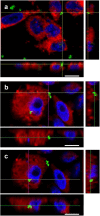
Figure 7. ACT is sufficient to promote internalisation of “toxin-coated beads”.
CHO-K1 cells were incubated with fluorescent “ACT-coated beads” (green fluorescence) and invasion assay and confocal microscopy analysis were performed as described in Experimental procedures. In control cells treated with BSA-coated beads the fluorescent beads were outside the cells (a) Analysis of the microscopy images revealed the presence of some fluorescent “ACT-coated” beads just “attached” or adhered to cell membranes (b) while other beads were clearly detected inside the cells (c). For Z-stack confocal analysis, cell membranes were stained with DiI (red) and DNA with Hoechst (blue). Representative images of three independent experiments are shown. Scale bars, 10 μm. A minimum of 30 cells were analyzed in each individual experiment.
Figure 8
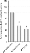
Figure 8. ACT-induced Ca2+ influx is necessary for the uptake of the “ACT-coated bacteria”.
CHO-K1 cells were pre-incubated for 30 min at 37 °C with nifedipine (10 μM), an inhibitor of L-type Ca2+ channels, or with KT5720 (56 nM), which specifically inhibits cAMP-dependent protein kinase A (involved in the activation of L-type channels), then the pretreated cells were exposed to “ACT-coated bacteria”, and the invasion assay was performed as described in Experimental procedures. The data were normalized to the control cells (no inhibitor addition) and expressed as per cent of control entry. Data shown are the mean ± SD of at least three independent experiments performed in quintuplicate, with *p < 0.01.

Similar articles

Cited by

References

    1. Guiso N. Bordetella pertussis: Why is it still circulating? J. Infect. 68, S119–S124 (2014). - PubMed
    1. Mooi F. R., Van Der Maas N. A. T. & De Melker H. E. Pertussis resurgence: Waning immunity and pathogen adaptation—Two sides of the same coin. Epidemiol. Infect. 142, 685–694 (2014). - PMC - PubMed
    1. de Gouw D., Diavatopoulos D. A., Bootsma H. J., Hermans P. W. & Mooi F. R. Pertussis: A matter of immune modulation. FEMS Microbiol. Rev. 35, 441–474 (2011). - PubMed
    1. Friedman R. L. Pertussis: The disease and new diagnostic methods. Clin. Microbiol. Rev. 1, 365–376 (1988). - PMC - PubMed
    1. Higgs R., Higgins S. C., Ross P. J. & Mills K. H. G. Immunity to the respiratory pathogen Bordetella pertussis. Mucosal Immunology 5, 485–500 (2012). - PubMed

Publication types

LinkOut - more resources