. 2015 Sep 8:5:13831.
doi: 10.1038/srep13831.

Microdissection of Human Esophagogastric Junction Wall with Phase-contrast X-ray CT Imaging

Affiliations

Microdissection of Human Esophagogastric Junction Wall with Phase-contrast X-ray CT Imaging

Jianfa Zhang et al. Sci Rep. .

Abstract

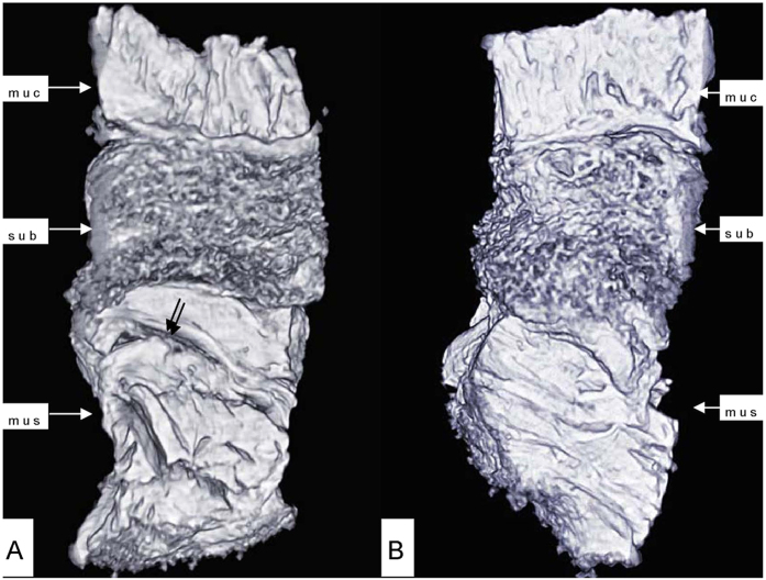
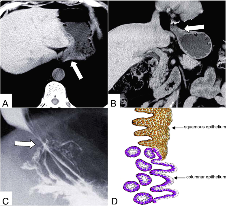
Phase-contrast x-ray imaging using an x-ray interferometer has great potential to reveal the structures inside soft tissues, because the sensitivity of this method to hydrogen, carbon, nitrogen, and oxygen is about 1000 times higher than that of the absorption-contrast x-ray method. In this study, we used phase-contrast X-ray CT to investigate human resected esophagogastric junction. This technology revealed the three-layer structure of the esophagogastric junction wall-mucous, submucosa and muscular layers. The mucous and muscular layers were clearly separated by a loose submucosa layer with a honeycomb appearance. The shape of the mucous and muscular layers was intact. The boundary between the mucous and submucosa layers was distinct, as was the border of the muscular and submucosa layers. The surface of the esophagogastric junction was displayed clearly through 3D reconstruction. The technology might be helpful in the diagnosis of esophagogastric junction lesion, especially for the early adenocarcinoma.


Figures

Figure 1
Figure 1. The picture of BL13W1 beamline partial facility of SSRF.
1. A multidimensional specimen table. The specimens are placed on the specimen table to rotate and specimens were obtained in different angles. 2. An X-ray CCD. It obtained specimens’ projective images with high-resolution. 3. The precise guide rail. It can control the exact distance from the CCD to the specimens.
Figure 2
Figure 2. Horizontal scan (A) and histology staining of the esophagogastric junction (B).
The normal esophagogastric junction wall was clearly depicted with Phase-contrast X-ray CT including the mucous, submucosa and muscular layers (A). muc: mucous layer; sub: submucosa layer; mus: muscular layer.
Figure 3
Figure 3. 3D image of the esophagogastric junction.
The shape of the mucous and muscular layers was complete. The boundary between the mucous and submucosa layers was distinct, and the same as the muscular and submucosa layers. The surface of the esophagogastric junction was displayed clearly. Crevice was revealed (double arrow in A).
Figure 4
Figure 4. Longitudinal scan of the esophagogastric junction.
Submucosa layer was displayed honeycomb appearance (A). The mucous and muscular layers were relatively close-grained (B,C). Some strips of tubular low-densuty areas in muscular layer were crevices (double arrow in C).
Figure 5
Figure 5
CT axial view image (A) coronal view image (B) Gas-barium double-contrast X-ray image (C) and sketch map (D) of the esophagogastric junction: (A,B) CT demonstrates the esophagogastric junction and the relation with adjacent tissues(the empty arrow). (C) Mucosa shows stellate high-density shadow (the empty arrow).

Similar articles

References

    1. Yassi R. et al. Three-dimensional high-resolution reconstruction of the human gastro-oesophageal junction. Clin. Anat. 23, 287–296 (2010). - PMC - PubMed
    1. Boyle J. T. et al. Role of the diaphragm in the genesis of lower esophageal sphincter pressure in the cat. Gastroenterology 88, 723–730 (1985). - PubMed
    1. McMahon B. P., Odie K. D., Moloney K. W. & Gregersen H. Computation of flow through the oesophagogastric junction. World J Gastroenterol 13, 1360–1364 (2007). - PMC - PubMed
    1. McMahon B. P., Drewes A. M. & Gregersen H. Functional oesophago-gastric junction imaging. World J Gastroenterol 12, 2818–2824 (2006). - PMC - PubMed
    1. Mittal R. K. & Balaban D. H. The esophagogastric junction. N. Engl. J. Med. 336, 924–932 (1997). - PubMed

Publication types

MeSH terms