. 2015 Sep 8;46(1):89.
doi: 10.1186/s13567-015-0244-6.

Piscine orthoreovirus (PRV) in red and melanised foci in white muscle of Atlantic salmon (Salmo salar)

Affiliations

Piscine orthoreovirus (PRV) in red and melanised foci in white muscle of Atlantic salmon (Salmo salar)

Håvard Bjørgen et al. Vet Res. .

Abstract

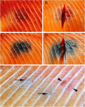
Melanised focal changes (black spots) are common findings in the white skeletal muscle of seawater-farmed Atlantic salmon (Salmo salar). Fillets with melanised focal changes are considered as lower quality and cause large economic losses. It has been suggested that red focal changes (red spots) precede the melanised focal changes. In the present work, we examined different populations of captive and wild salmon for the occurrence of both types of changes, which were investigated for the presence of different viruses by immunohistochemistry and RT-qPCR. The occurrence of red or melanised foci varied significantly between the populations, from none in wild fish control group, low prevalence of small foci in fish kept in in-house tanks, to high prevalence of large foci in farm-raised salmon. Large amounts of Piscine orthoreovirus (PRV) antigen were detected in all foci. No other viruses were detected. Red focal changes contained significantly higher levels of PRV RNA than apparently non-affected areas in white muscle of the same individuals. Some changes displayed a transient form between a red and melanised pathotype, indicating a progression from an acute to a chronic manifestation. We conclude that PRV is associated with the focal pathological changes in the white muscle of farmed Atlantic salmon and is a premise for the development of focal melanised changes.


Figures

Figure 1
Figure 1
Gross pathological changes of white muscle. (A) A red focal change in the muscle of the craniodorsal region of the abdominal wall. (B) An incision through a red focal change showing discolouration extending deep into the fillet. (C) A melanised focal change detected in the same anatomical location in a different fish. (D) A melanised focal change with extensive amounts of a whitish tissue extending into the depth of the fillet. (E) A muscle fillet with two faint melanised lesions (arrows) and one red focal change (arrowheads).
Figure 2
Figure 2
Gross image of paraffin-embedded red focal change. Transient form with both red (asterisk) and melanised (arrowheads) changes. Scale bar = 1 cm.
Figure 3
Figure 3
Histological investigations of red focal changes. (A) Necrotic myocytes display pale cytoplasm and appear homogenous without striations (arrow). Myocyte with striations (asterisk) (HE staining). (B) Necrotic myocytes with erythrocytes apparently located within the myocytes (arrowheads) and some infiltrations of macrophage-like cells (arrow) (HE staining). (C) Centrally located vacuoles of varying size, surrounded by moderate levels of collagen (intense red) and modest numbers of leukocytes (van Gieson staining). (D) Focal haemorrhage surrounding vacuoles. The edges of the vacuoles are positive for ferric iron (blue colour) (Perls’ Berlin blue staining). (A, C) scale bar = 200 μm, (B) scale bar = 100 μm, (D) scale bar = 50 μm.
Figure 4
Figure 4
Histological investigations of melanised focal changes. (A) Transverse section of necrotic muscle cells (asterisk) and severe fibrosis (arrow) with infiltrates of leukocytes. A cell-rich granuloma with melanin-containing cells is present in the right part of the picture (G), displaying a heterogeneous morphology (HE staining). (B) Severe vacuolisation and adipocytes with surrounding melano-macrophages (black) and clusters of regenerating myocytes with a basophilc cytoplasm (arrow) (HE staining). (C) Multiple vacuoles in an area with severe fibrosis (intense red staining) (van Gieson staining). (D) Several iron-containing macrophage-like cells (blue staining) in association with melano-macrophages (black) (Pearls’ Berlin blue staining). (A, C) scale bar = 100 μm, (B, D) scale bar = 50 μm.
Figure 5
Figure 5
Histological image of cardiac spongious layer. Infiltrates of leukocytes are visible in endocard and myocard. Scale bar = 100 μm.
Figure 6
Figure 6
Immunohistochemical staining for PRV antigens of red (A-B) and melanised (C-H) focal changes. (A) Abundant amounts of PRV-positive cells (brown) and erythrocytes in the necrotic muscle tissue. (B) Transverse section of necrotic myocytes with intracellular PRV-positive macrophage-like cells and erythrocytes (brown). Distinct granular staining is present in the cytoplasm of the macrophage-like cells. (C) A well-organised granuloma is present in the center of the image, heavily positive for PRV (red), and with vast amounts of elongated melano-macrophages (black). (D) A single granuloma with PRV-positive cells (red) surrounded by heavily pigmented melano-macrophages. (E) A focus with necrotic tissue and infiltrates of melano-macrophages (black) and macrophage-like cells positive for PRV (red). (F) A close-up of E where the distinct reaction in the cytoplasm is evident (red). (G) PRV antigen (red) in melano-macrophages (black) in a necrotic myocyte. Higher resolution image in the upper right corner (100 x). (H) A single necrotic myocyte undergoing phagocytosis and containing abundant PRV-positive macrophage-like cells (red). (A, C) scale bar = 200 μm, (E) scale bar = 100 μm, (B, D, F, G, H) scale bar = 50 μm
Figure 7
Figure 7
RT-qPCR analysis for PRV in blood (A) and white skeletal muscle (B). Fish groups with melanised, red or no changes are coloured brown, red, and grey, respectively. (A) Detection in blood presented by box plot with whiskers indicating max/min of the Ct-values from Group A (n = 25), B (n = 35), E (n = 20), F (n = 42), G (n = 43) and H (n = 10). The percent of fish in each group with melanised focal changes are displayed. (B) Samples from red or melanised focal changes (+) and non-affected areas (−) from the same individual. Box plot and whiskers showing max/min of the Ct-values from Group C with red focal changes (n = 6), and from Groups A (n = 9) and E (n = 14) with melanised focal changes. Only non-affected tissue (−) could be tested from groups without changes; i.e. Groups F (n = 6) and G (n = 6). Analyses were performed using Wilcoxon matched pairs signed rank test. *p < 0.05.

References

    1. Koppang EO, Haugarvoll E, Hordvik I, Aune L, Poppe TT (2005) Vaccine-associated granulomatous inflammation and melanin accumulation in Atlantic salmon, Salmo salar L. white muscle. J Fish Dis 28:13–22 - PubMed
    1. Mørkøre T, Heia K (2012) Black spots in salmon fillet - extent and methods of measurement. Norsk fiskeoppdrett 3:50–53
    1. Larsen HA, Austbø L, Mørkøre T, Thorsen J, Hordvik I, Fischer U, Jirillo E, Rimstad E, Koppang EO. Pigment-producing granulomatous myopathy in Atlantic salmon: a novel inflammatory response. Fish Shellfish Immunol. 2012;33:277–285. doi: 10.1016/j.fsi.2012.05.012. - DOI - PubMed
    1. Roberts RJ, McQueen A, Shearer WM, Young H. The histopathology of salmon tagging. J Fish Biol. 1973;5:615–619. doi: 10.1111/j.1095-8649.1973.tb04495.x. - DOI
    1. Haugarvoll E, Thorsen J, Laane M, Huang Q, Koppang EO. Melanogenesis and evidence for melanosome transport to the plasma membrane in a CD83 teleost leukocyte cell line. Pigment Cell Res. 2006;19:214–225. doi: 10.1111/j.1600-0749.2006.00297.x. - DOI - PubMed

LinkOut - more resources