. 2015 Sep 8:5:13834.
doi: 10.1038/srep13834.

Modeling the interplay between the HIF-1 and p53 pathways in hypoxia

Affiliations

Modeling the interplay between the HIF-1 and p53 pathways in hypoxia

Chun-Hong Zhou et al. Sci Rep. .

Abstract

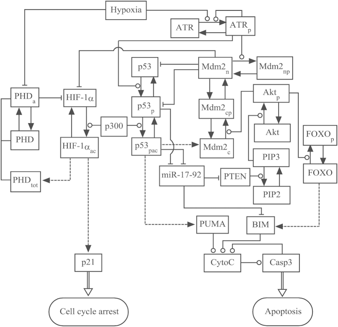
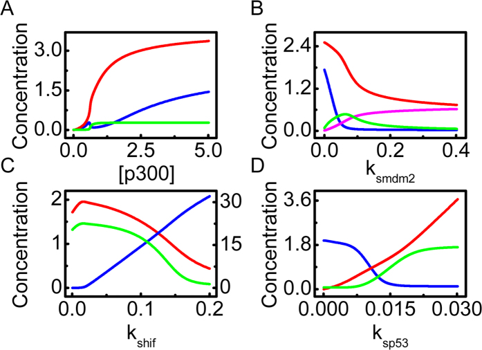
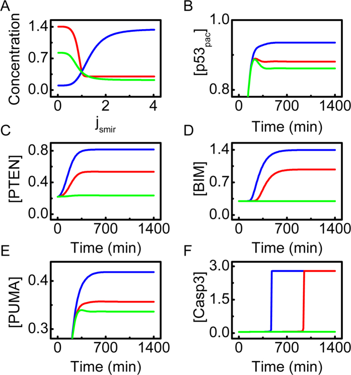
Both the hypoxia-inducible factor-1 (HIF-1) and tumor suppressor p53 are involved in the cellular response to hypoxia. How the two transcription factors interact to determine cell fates is less well understood. Here, we developed a network model to characterize crosstalk between the HIF-1 and p53 pathways, taking into account that HIF-1α and p53 are targeted for proteasomal degradation by Mdm2 and compete for binding to limiting co-activator p300. We reported the network dynamics under various hypoxic conditions and revealed how the stabilization and transcriptional activities of p53 and HIF-1α are modulated to determine the cell fate. We showed that both the transrepression and transactivation activities of p53 promote apoptosis induction. This work provides new insight into the mechanism for the cellular response to hypoxia.


Figures

Figure 1
Figure 1. Schematic depiction of the model.
The model characterizes crosstalk between the HIF-1 and p53 pathways upon hypoxia. In hypoxia, HIF-1α is stabilized due to reduced hydroxylation by PHD. Under severe hypoxia, the ATR kinase is activated via auto-phosphorylation upon hypoxia-induced replication arrest, and p53 is further activated by ATR. The shared coactivator p300 is required for the full transcriptional activity of p53 and HIF-1α. HIF-1α evokes transient cell-cycle arrest via inducing p21, whereas p53 can induce apoptosis via transrepressing or/and transactivating target genes. Dashed lines denote the expression of target genes by HIF-1α or p53, while solid arrowed lines represent the transitions between proteins. Circle- and bar-headed lines denote the promotion and inhibition of transition or production, respectively.
Figure 2
Figure 2. Overview of the network dynamics under different hypoxic conditions.
Temporal evolution of the levels of ATRp, p53pac, HIF-1αac, p21 and Casp3 in mild hypoxia (2% O2, (A)), severe hypoxia (0.02% O2, (B)), or anoxia (0% O2, (C)).
Figure 3
Figure 3. Dynamics of HIF-1αac and p21.
Color-coded concentrations of (A) HIF-1αac and (B) p21 as a function of the logarithm of oxygen concentration and time.
Figure 4
Figure 4. Protein dynamics and apoptosis induction under severe hypoxia.
(A–D) Color-coded concentrations of ATRp (A), p53pac (B), BIM (C) and PUMA (D) as a function of oxygen concentration and time. (E) The timing of Casp3 activation versus oxygen concentration. (F) Time courses of [Casp3] with oxygen concentration at 0.01% (green), 0.018% (blue), or 0.02% (red).
Figure 5
Figure 5. Interplay between HIF-1α and p53 in anoxia.
(A) Bifurcation diagrams of [p53pac] (red), [HIF-1αac] (blue) and [Mdm2n] (green) versus the amount of p300. (B) Bifurcation diagrams of [p53pac] (red), [HIF-1αac] (blue), [Mdm2c] (green) and [Mdm2n] (pink) versus the p53-induced production rate of Mdm2, ksmdm2. (C) Bifurcation diagrams of [HIF-1αac] (blue, on the right axis), [p53pac] (red) and [PUMA] (green) versus the production rate of HIF-1α, kshif. (D) Bifurcation diagrams of [HIF-1αac] (blue), [p53pac] (red) and [PUMA] (green) versus the production rate of p53, ksp53.
Figure 6
Figure 6. Effect of miR-17-92 on apoptosis induction under severe hypoxia (0.02% O2).
(A) Bifurcation diagrams of [PTEN] (green), [BIM] (red) and [miR-17-92] (blue) as a function of jsmir. (BF) Time courses of [p53pac] (B), [PTEN] (C), [BIM] (D), [PUMA] (E) and [Casp3] (F) with jsmir = 0.3 (blue), 0.8 (red), or 2.0 (green).

Similar articles

Cited by

References

    1. Semenza G. L. HIF-1: Upstream and downstream of cancer metabolism. Curr. Opin. Genet. Dev. 20, 51–56 (2010). - PMC - PubMed
    1. Wang G. L., Jiang B. H., Rue E. A. & Semenza G. L. Hypoxia-inducible factor 1 is a basic-helix-loop-helix-PAS heterodimer regulated by cellular O2 tension. Proc. Natl. Acad. Sci. USA 92, 5510–5514 (1995). - PMC - PubMed
    1. Ivan M. et al. HIFα targeted for VHL-mediated destruction by proline hydroxylation: Implications for O2 sensing. Science 292, 464–468 (2001). - PubMed
    1. Lando D. et al. FIH-1 is an asparaginyl hydroxylase enzyme that regulates the transcriptional activity of hypoxia-inducible factor. Genes Dev. 16, 1466–1471 (2002). - PMC - PubMed
    1. Semenza G. L. Targeting HIF-1 for cancer therapy. Nat. Rev. Cancer 3, 721–732 (2003). - PubMed

Publication types

MeSH terms

Substances