

Radiographic development during three decades in a patient with psoriatic arthritis mutilans
- PMID: 26346445
- PMCID: PMC4548743
- DOI: 10.1177/2058460115588098
Radiographic development during three decades in a patient with psoriatic arthritis mutilans
Abstract
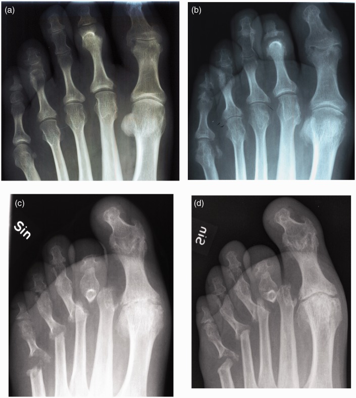
Psoriatic arthritis mutilans (PAM) is the most severe and rare form of psoriatic arthritis (PsA). We describe radiological development in a typical case of PAM covering three decades in order to elucidate the need for early diagnosis of PAM. Radiographs of hands and feet, taken from 1981 to 2010, were evaluated using the Psoriatic Arthritis Ratingen Score (PARS). When PsA was diagnosed, in 1981, gross deformity was observed in the second PIP joint of the left foot. Several pencil-in-cup deformities and gross osteolysis were present in the feet in the first decade of the disease. Over 10 years, many joints had reached maximum scores. During the follow-up, other joints became involved and the disease developed clinically. Reporting early signs suggestive of PAM, e.g. pencil-in cup deformities and gross osteolysis in any joint, should be mandatory and crucial. This would heighten our awareness of PAM, accelerate the diagnosis, and lead to improved effective treatment in order to minimize joint damages resulting in PAM.
Keywords: Radiology; arthritis; follow-up; mutilans; psoriasis.
Figures

Similar articles
-
Radiographic scoring systems for psoriatic arthritis are insufficient for psoriatic arthritis mutilans: results from the Nordic PAM Study.Acta Radiol Open. 2020 Apr 27;9(4):2058460120920797. doi: 10.1177/2058460120920797. eCollection 2020 Apr. Acta Radiol Open. 2020. PMID: 32426163 Free PMC article.
-
Psoriatic Arthritis Mutilans: Characteristics and Natural Radiographic History.J Rheumatol. 2015 Jul;42(7):1169-76. doi: 10.3899/jrheum.150083. Epub 2015 May 15. J Rheumatol. 2015. PMID: 25979723
-
Follow-up of psoriatic arthritis mutilans patients treated with anti-TNF-alpha therapy.J Drugs Dermatol. 2009 Apr;8(4):406-12. J Drugs Dermatol. 2009. PMID: 19363860
-
Arthritis mutilans.Curr Rheumatol Rep. 2013 Apr;15(4):321. doi: 10.1007/s11926-013-0321-7. Curr Rheumatol Rep. 2013. PMID: 23430715 Review.
-
Radiographic scoring methods in psoriatic arthritis.Clin Exp Rheumatol. 2015 Sep-Oct;33(5 Suppl 93):S55-9. Epub 2015 Oct 15. Clin Exp Rheumatol. 2015. PMID: 26472605 Review.
Cited by
-
Radiographic scoring systems for psoriatic arthritis are insufficient for psoriatic arthritis mutilans: results from the Nordic PAM Study.Acta Radiol Open. 2020 Apr 27;9(4):2058460120920797. doi: 10.1177/2058460120920797. eCollection 2020 Apr. Acta Radiol Open. 2020. PMID: 32426163 Free PMC article.
-
Comorbidities in a Cohort of 66 Patients With Psoriatic Arthritis Mutilans-Results From the Nordic PAM Study.Front Med (Lausanne). 2021 Feb 4;8:629741. doi: 10.3389/fmed.2021.629741. eCollection 2021. Front Med (Lausanne). 2021. PMID: 33614686 Free PMC article.
-
Rare coding variants in NOX4 link high ROS levels to psoriatic arthritis mutilans.EMBO Mol Med. 2024 Mar;16(3):596-615. doi: 10.1038/s44321-024-00035-z. Epub 2024 Feb 20. EMBO Mol Med. 2024. PMID: 38379095 Free PMC article.
References
-
- Moll JM, Wright V. Psoriatic arthritis. Semin Arthritis Rheum 1973; 3: 55–78. - PubMed
-
- Haddad A, Chandran V. Arthritis mutilans. Curr Rheumatol Rep 2013; 15: 321–326. - PubMed
-
- Tam LS, Leung YY, Li EK. Psoriatic arthritis in Asia. Rheumatology (Oxford) 2009; 48: 1473–1477. - PubMed
-
- Scarpa R. Clinical manifestation and diagnosis of psoriatic arthritis. Rheumatology in Europe 1998; 27: 130–132.
-
- Koo T, Nagy Z, Sesztak M, et al. Subsets in psoriatic arthritis by cluster analysis. Clin Rheumatol 2001; 20: 36–43. - PubMed
Publication types
LinkOut - more resources
Full Text Sources
Other Literature Sources
Research Materials
Miscellaneous
