Review
. 2015 Dec;72(23):4593-612.
doi: 10.1007/s00018-015-2031-y. Epub 2015 Sep 7.

Role of the nucleus in apoptosis: signaling and execution

Affiliations
Review

Role of the nucleus in apoptosis: signaling and execution

Evgeniia A Prokhorova et al. Cell Mol Life Sci. 2015 Dec.

Abstract

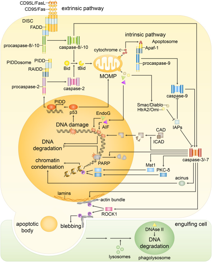
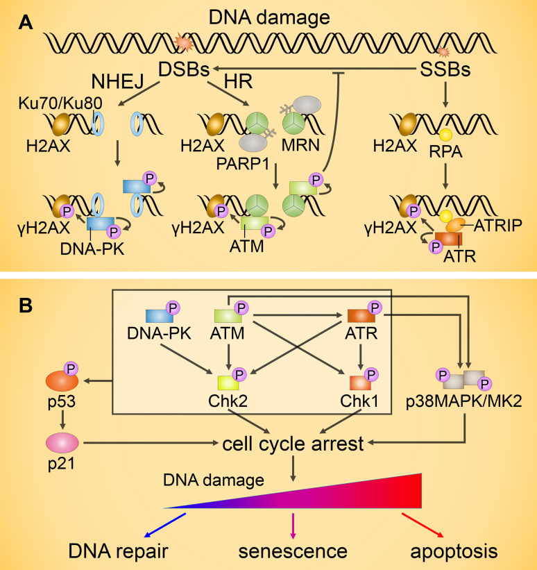
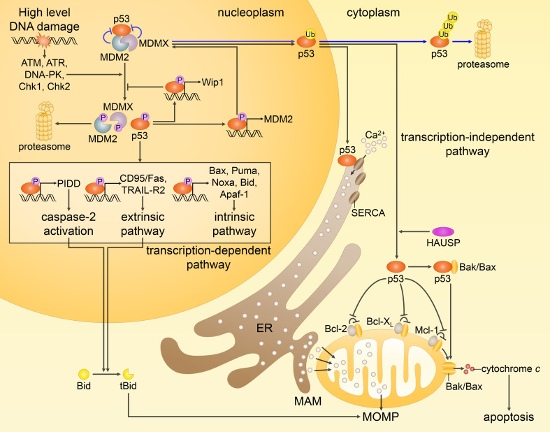
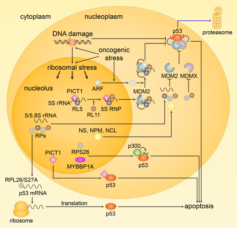
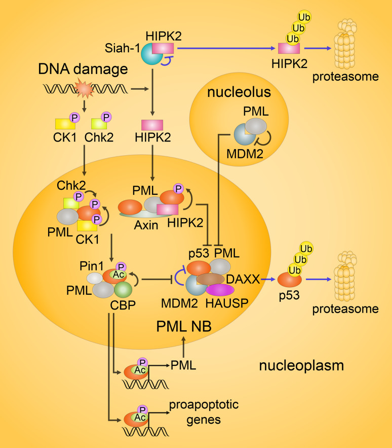
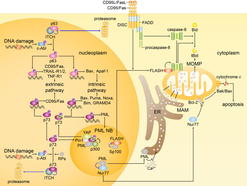
Since their establishment in the early 1970s, the nuclear changes upon apoptosis induction, such as the condensation of chromatin, disassembly of nuclear scaffold proteins and degradation of DNA, were, and still are, considered as the essential steps and hallmarks of apoptosis. These are the characteristics of the execution phase of apoptotic cell death. In addition, accumulating data clearly show that some nuclear events can lead to the induction of apoptosis. In particular, if DNA lesions resulting from deregulation during the cell cycle or DNA damage induced by chemotherapeutic drugs or viral infection cannot be efficiently eliminated, apoptotic mechanisms, which enable cellular transformation to be avoided, are activated in the nucleus. The functional heterogeneity of the nuclear organization allows the tight regulation of these signaling events that involve the movement of various nuclear proteins to other intracellular compartments (and vice versa) to initiate and govern apoptosis. Here, we discuss how these events are coordinated to execute apoptotic cell death.

Keywords: Caspases; Endonucleases; PML nuclear bodies; Ribosomal stress; p53; p63; p73.


Figures

Fig. 1
Fig. 1
Both extrinsic and intrinsic apoptotic signaling pathways result in the same changes in the nuclear morphology. The extrinsic pathway begins with the stimulation of the death receptors by their cognate ligands that results in the assembly of initiator caspase-8/-10 activation platforms, such as death-inducing signaling complex (DISC). Caspase-8/-10 activation is followed by the cleavage of effector caspase-3, -6 and -7 and, in some cases, processing of Bid: Truncated Bid (tBid) subsequently leads to outer mitochondrial membrane permeabilization (MOMP), thereby, engaging the intrinsic pathway. The major trigger of the intrinsic pathway is DNA damage, following which MOMP is induced via several transcriptional and non-transcriptional mechanisms. MOMP results in the efflux of cytochrome c, which engages the apoptosome formation required for the activation of caspase-9. The mitochondrial proteins Smac/DIABLO and HtrA2/Omi promote apoptosis by neutralizing inhibitors of apoptosis proteins (IAPs), thus reversing their grip on caspase-3, -7, -9. In addition, in response to DNA damage formation of PIDDosome, a platform for caspase-2 activation, might be engaged. Initiator caspase-2 and -9, as well as -8 and -10, propagate the apoptotic signal by direct cleavage of the effector caspases. Effector caspases coordinate the dismantling of the nucleus. Caspase-mediated cleavage of poly(ADP-ribose) polymerase (PARP) prevents DNA repair and facilitates access of nucleases to the chromatin. Then, cleavage of inhibitor of caspase-activated DNase (ICAD) releases CAD, which enters the nucleus and catalyzes DNA degradation. Meanwhile, DNA fragmentation is also promoted in a caspase-independent manner by endonuclease G (EndoG) and apoptosis-inducing factor (AIF), which are released upon MOMP. Caspase-dependent activation of Mst1, PKC-δ and acinus stimulates chromatin condensation. Caspase-mediated cleavage of nuclear lamins contributes to nuclear fragmentation, whereas the proteolysis of the Rho effector ROCK1 results in contraction of the actin cytoskeleton that promotes breakdown of the nuclear envelope, as well as plasma membrane blebbing. Eventually, the cell collapses into apoptotic bodies, which are rapidly engulfed by phagocytic cells. Subsequently, DNA fragments, which were kept within apoptotic bodies, are digested by phagocytic DNase II
Fig. 2
Fig. 2
Transduction of DNA damage signals. a Activation of ataxia-telangiectasia mutated (ATM), Rad3-related (ATR) and DNA–protein kinase (DNA-PK) upon DNA damage. When a double-strand break (DSB) occurs, it is generally repaired via the non-homologous end-joining (NHEJ) or homologous recombination (HR) pathways. NHEJ is engaged upon Ku70/Ku80 binding to DSBs followed by recruitment and activation of DNA-PK. During HR, PARP1 binds to the DSBs and mediates the initial recruitment of the MRE11/Rad50/NBS1 (MRN) complex and subsequent binding and activation of ATM. Single-strand breaks (SSBs) repair is initiated by replication protein A (RPA), which recruits the ATR/ATR-interacting protein (ATRIP) complex and results in ATR activation. Activation of ATM, ATR, and DNA-PK is followed by the phosphorylation of H2AX that amplifies the molecular signal by DNA repair protein recruitment. b ATM, ATR, and DNA-PK can phosphorylate and activate the transcription factor p53 either directly or by means of prior activation of checkpoint kinases Chk1/2. Both ATM and ATR also contribute to the activation of the p38MAPK/MK2 kinase complex. Activation of Chk1/2 and MK2 downstream targets, as well as p53-mediated upregulation of the Cdk2 inhibitor p21, results in transient cell cycle arrest. Unrepaired DNA damage generally leads to permanent cell cycle arrest (senescence) or apoptosis
Fig. 3
Fig. 3
Different routes of p53-mediated apoptotic cell death. In response to extensive DNA damage, the interactions between p53, MDM2 and MDMX are disrupted by posttranslational modifications (only some phosphorylation events are shown), preventing inhibition and proteasomal degradation of p53 (p53 is normally kept at low levels by MDM2 and MDMX, as represented by the blue arrows). Activated p53 upregulates proapoptotic genes (as highlighted in the box), and represses antiapoptotic genes (not shown). As a result, p53 predominantly engages the intrinsic apoptotic pathway. In addition, p53 transactivates MDM2 and Wip1 phosphatase, providing the negative feedback loops that help to restrain p53 at the end of a stress response. Non-transcriptional activities of p53, as shown on the right, include translocation of its monoubiquitinated form to the mitochondria, where, after being deubiquitinated by HAUSP, it promotes Bak/Bax oligomerization through direct physical interaction with Bak/Bax or by binding of antiapoptotic proteins Bcl-2, Bcl-XL and Mcl-1. On the other hand, cytosolic p53 interacts with the sarco-ER Ca2+-ATPase (SERCA) pumps at the endoplasmic reticulum (ER) and mitochondrial-associated membranes (MAMs), potentiating Ca2+ influx followed by an enhanced transfer to the mitochondria. These transcription-independent functions of p53, along with the transcription-mediated events, prompt MOMP leading to the release of cytochrome c and subsequent apoptosis
Fig. 4
Fig. 4
Schematic representation of the ribosomal (nucleolar) stress response. Under normal conditions, ribosome biogenesis occurs normally and p53 activity is kept low (indicated by the blue arrows). Following ribosomal stress, ribosome biogenesis is perturbed, and nucleolar proteins, including rRNAs and ribosomal proteins (RPs), are released into the nucleoplasm leading to the activation of p53. 5S RNP, consisting of  RPL5, RPL11 and 5S rRNA, seems to be the main player in eliciting p53 response upon impairment of ribosomal biogenesis; it acts via direct binding and inhibition of MDM2, and is essential for the sequestration of MDM2 by ARF. Along with 5S RNP and ARF, free rRNAs and RPs, and other nucleolar proteins [e.g., nucleostemin (NS), nucleophosmin (NPM) and nucleolin (NCL)] also contribute to MDM2 inhibition by binding, while a number of RPs act through the inhibition of MDMX or upregulation of p53 mRNA translation (RPL26 and RPS27A). In addition, RPS26 and MYBBP1A promote p53-mediated transcription through the facilitation of p53 acetylation by p300. The role of PICT1 remains controversial, but since, for instance, it facilitates the formation of 5S RNP and can directly increase p53 activity by binding, it appears to be an important mediator of the ribosomal stress response
Fig. 5
Fig. 5
Regulation of p53 activity in the promyelocytic leukemia nuclear bodies (PML NBs). In response to DNA damage, several kinases, including Chk2 and CK1, as well as HIPK2 in lethally damaged cells, are activated. Under unstressed or mildly stressed conditions, HIPK2 is targeted for proteasomal degradation (the blue arrows on the top). MDM2-mediated ubiquitination of p53 in unstressed cells is promoted by DAXX and HAUSP, as indicated by the blue arrows in the middle. Following DNA damage, phosphorylation of p53 by Chk2, CK1 and HIPK2 in PML NBs disrupts p53/MDM2 interaction. In addition, PML inhibits MDM2 by sequestering it in the nucleoli. Moreover, upon severe genotoxic stress, Axin, DAXX or Tip60 (not shown) might form complexes with HIPK2 and p53, enhancing the function of HIPK2 against p53. In turn, the phosphorylation of p53 drives its acetylation. In PML NBs, acetylation is mainly performed by CBP, while Pin1 promotes this  event. Activated p53 induces the expression of its target genes, including PML
Fig. 6
Fig. 6
p63, p73, FLASH and Nur77 as initiators of p53-independent apoptosis. In response to DNA damage, c-Abl kinase is activated and accumulated in the nucleus. Subsequently, p63 and p73 are phosphorylated, and their targeting for proteasomal degradation by ITCH (blue arrows) is prevented. Moreover, p73 is recruited to PML NBs, where it is acetylated by p300 (and to the lesser extent by pCAF), while YAP and Pin1 promote the modification. In addition, such RPs as RPL5, RPL11 and RPS14 bind to p73 enhancing its transcriptional activity. Activated p63 and p73 induce the expression of several proapoptotic genes engaging the intrinsic and extrinsic apoptotic pathways. At the same time, p73 might move to the cytoplasm and promote MOMP independently of its transcriptional activity (not shown). In unstressed cells, FLASH colocalizes with Sp100 in PML NBs. Following CD95/Fas activation, FLASH relocates to the cytosol and accumulates at the mitochondria, where it binds caspase-8, and promotes its activation followed by Bid cleavage. Nur77, when in the nucleus, acts as a transcription factor that promotes the expression of prosurvival factors. In response to stress stimuli, Nur77 translocates to the cytoplasm and reverses antiapoptotic function of Bcl-2 promoting the formation of Bak/Bax channels that results in MOMP. Translocation of nuclear PML to the cytoplasm may lead to its accumulation at the ER and MAMs and subsequent upregulation of the Ca2+ transfer to the mitochondria followed by the loss of membrane potential and apoptosis

Similar articles

Cited by

References

    1. Kerr JFR, Wyllie AH, Currie ARD. Apoptosis: a basic biological phenomenon with wide-ranging implications in tissue kinetics. Br J Cancer. 1972;26:239–257. doi: 10.1038/bjc.1972.33. - DOI - PMC - PubMed
    1. Skalka M, Matyasova J, Cejkova M. DNA in chromatin of irradiated lymphoid tissues degrades in vivo into regular fragments. FEBS Lett. 1976;72:271–275. doi: 10.1016/0014-5793(76)80984-2. - DOI - PubMed
    1. Wyllie AH. Glucocorticoid-induced thymocyte apoptosis is associated with endogenous endonuclease activation. Nature. 1980;284:555–556. doi: 10.1038/284555a0. - DOI - PubMed
    1. Ellis HM, Horvitz HR. Genetic control of programmed cell death in the nematode C. elegans . Cell. 1986;44:817–829. doi: 10.1016/0092-8674(86)90004-8. - DOI - PubMed
    1. Jacobson MD, Burne JF, Raff MC. Programmed cell death and Bcl-2 protection in the absence of a nucleus. EMBO J. 1994;13:1899–1910. - PMC - PubMed

Publication types