

A Method to Improve Electron Density Measurement of Cone-Beam CT Using Dual Energy Technique
- PMID: 26346510
- PMCID: PMC4540959
- DOI: 10.1155/2015/858907
A Method to Improve Electron Density Measurement of Cone-Beam CT Using Dual Energy Technique
Abstract
Purpose: To develop a dual energy imaging method to improve the accuracy of electron density measurement with a cone-beam CT (CBCT) device.
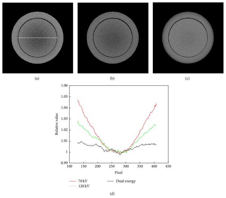
Materials and methods: The imaging system is the XVI CBCT system on Elekta Synergy linac. Projection data were acquired with the high and low energy X-ray, respectively, to set up a basis material decomposition model. Virtual phantom simulation and phantoms experiments were carried out for quantitative evaluation of the method. Phantoms were also scanned twice with the high and low energy X-ray, respectively. The data were decomposed into projections of the two basis material coefficients according to the model set up earlier. The two sets of decomposed projections were used to reconstruct CBCT images of the basis material coefficients. Then, the images of electron densities were calculated with these CBCT images.
Results: The difference between the calculated and theoretical values was within 2% and the correlation coefficient of them was about 1.0. The dual energy imaging method obtained more accurate electron density values and reduced the beam hardening artifacts obviously.
Conclusion: A novel dual energy CBCT imaging method to calculate the electron densities was developed. It can acquire more accurate values and provide a platform potentially for dose calculation.
Figures


Similar articles
-
Dual-energy imaging method to improve the image quality and the accuracy of dose calculation for cone-beam computed tomography.Phys Med. 2017 Apr;36:110-118. doi: 10.1016/j.ejmp.2017.03.023. Epub 2017 Apr 4. Phys Med. 2017. PMID: 28410679
-
Implementation of dual-energy technique for virtual monochromatic and linearly mixed CBCTs.Med Phys. 2012 Oct;39(10):6056-64. doi: 10.1118/1.4752212. Med Phys. 2012. PMID: 23039644
-
Combining scatter reduction and correction to improve image quality in cone-beam computed tomography (CBCT).Med Phys. 2010 Nov;37(11):5634-44. doi: 10.1118/1.3497272. Med Phys. 2010. PMID: 21158275
-
Generation of virtual monochromatic CBCT from dual kV∕MV beam projections.Med Phys. 2013 Dec;40(12):121910. doi: 10.1118/1.4824324. Med Phys. 2013. PMID: 24320521
-
Optimizing dual energy cone beam CT protocols for preclinical imaging and radiation research.Br J Radiol. 2017 Jan;90(1069):20160480. doi: 10.1259/bjr.20160480. Epub 2016 Nov 2. Br J Radiol. 2017. PMID: 27683003 Free PMC article. Review.
Cited by
-
Fast-switching dual energy cone beam computed tomography using the on-board imager of a commercial linear accelerator.Phys Med Biol. 2020 Jan 13;65(1):015013. doi: 10.1088/1361-6560/ab5c35. Phys Med Biol. 2020. PMID: 31775131 Free PMC article.
-
Fast, automated optimization of virtual monoenergetic images with the dual-energy image synthesizer for cone-beam CT.J Appl Clin Med Phys. 2025 Jun;26(6):e70083. doi: 10.1002/acm2.70083. Epub 2025 Apr 22. J Appl Clin Med Phys. 2025. PMID: 40260755 Free PMC article.
-
Technical note: TIGRE-DE for the creation of virtual monoenergetic images from dual-energy cone-beam CT.Med Phys. 2024 Apr;51(4):2975-2982. doi: 10.1002/mp.17002. Epub 2024 Feb 26. Med Phys. 2024. PMID: 38408013 Free PMC article.
References
-
- Saito T., Morohashi H., Hasebe T., et al. A review of stereotactic radiotherapy (SRT) for lung metastasis of colon cancer. Gan To Kagaku Ryoho. 2014;41(12):1462–1464. - PubMed
MeSH terms
LinkOut - more resources
Full Text Sources
Other Literature Sources
