. 2015 Sep 8:5:13841.
doi: 10.1038/srep13841.

Dimerization of lipocalin allergens

Affiliations

Dimerization of lipocalin allergens

Merja H Niemi et al. Sci Rep. .

Abstract

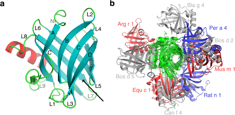
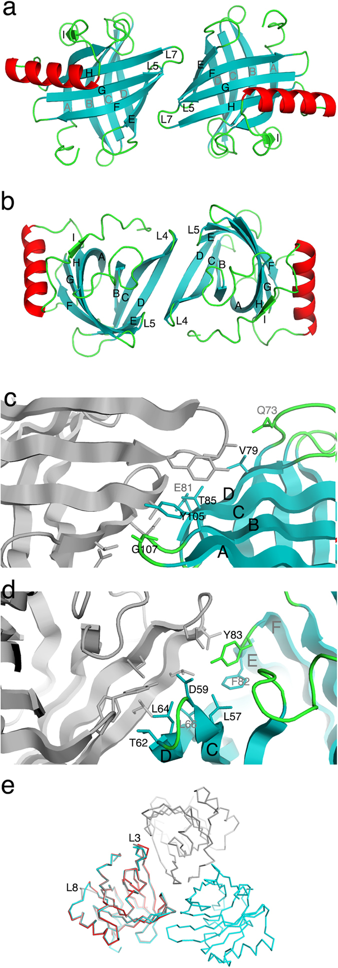
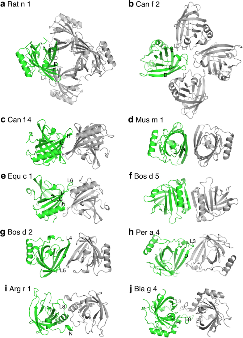
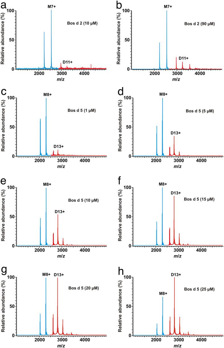
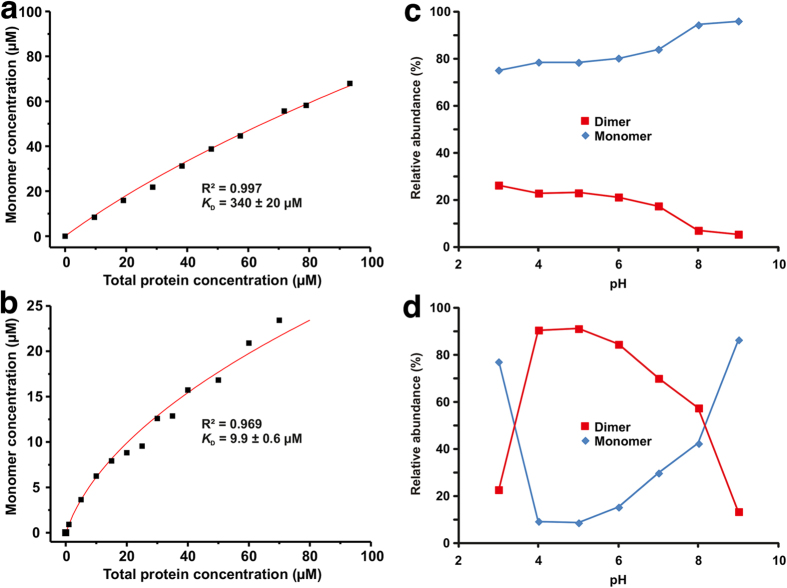
Lipocalins are one of the most important groups of inhalant animal allergens. The analysis of structural features of these proteins is important to get insights into their allergenicity. We have determined two different dimeric crystal structures for bovine dander lipocalin Bos d 2, which was earlier described as a monomeric allergen. The crystal structure analysis of all other determined lipocalin allergens also revealed oligomeric structures which broadly utilize inherent structural features of the β-sheet in dimer formation. According to the moderate size of monomer-monomer interfaces, most of these dimers would be transient in solution. Native mass spectrometry was employed to characterize quantitatively transient dimerization of two lipocalin allergens, Bos d 2 and Bos d 5, in solution.


Conflict of interest statement

M.N., J.J. and J.R. are shareholders of Desentum Oy. The rest of the authors declare that they have no relevant conflicts of interest.

Figures

Figure 1
Figure 1
(a) The ribbon representation for monomeric Bos d 2. The eight antiparallel β-strands (AH) forming a central barrel are shown as cyan arrows. The β-strand (I) outside the barrel is in green. The α-helix is in red and loops connecting different secondary structure elements are labelled L1 to L8. The black arrow shows the entrance to the ligand binding pocket (b) The monomers of 9 different lipocalin allergens which form symmetric homodimers are superimposed to observe the orientation of the second monomer. The superimposed monomers are in green and grey, blue and red colours are used for the second monomers.
Figure 2
Figure 2. Dimerization of Bos d 2 observed in crystals as a ribbon presentation.
(a) The symmetric dimer of Bos d 2 observed in the monoclinic (C2) crystal form, (b) in the trigonal (P3221) crystal form. The key residues which contribute to the binding on the monomer-monomer interface in Bos d 2 dimer in monoclinic (c) and in trigonal (d) crystal forms. (e) The superimposition of a Cα-backbone of monomeric Bos d 2 (orthorhombic, in red), dimeric Bos d 2 (monoclinic, in cyan), and dimeric Bos d 2 (trigonal, in grey).
Figure 3
Figure 3. The oligomeric structures for 10 lipocalin allergens observed in crystals.
One monomer is in green, other monomers in grey. The structural elements forming monomer-monomer interfaces are labelled.
Figure 4
Figure 4. Native mass spectra of lipocalin allergens at different protein concentrations.
(a,b) rBos d 2 at protein (monomer) concentrations of 10 and 90 μM. The peaks representing the protein monomer (M) are in blue, while the peaks representing the protein dimer (D) are in red. The most abundant peaks for the monomer and the dimer have been assigned with the number indicating the ion charge state (i.e., M7+ = [rBos d 2 + 7H]7+). (c-h) Native mass spectra of Bos d 5 at a protein (monomer) concentration of 1-25 μM.
Figure 5
Figure 5. Protein monomer concentration as a function of total protein concentration for (a) rBos d 2 and (b) Bos d 5 as calculated from the native mass spectra.
The solid lines are the best fits of the data to Equation 4 (see, Online methods for details) for determining KD value for dimerization. Relative abundances of the protein monomer and the dimer for (c) rBos d 2 and (d) Bos d 5 depending on solution pH at a protein (monomer) concentration of 40 μM.
Figure 6
Figure 6. The sketch for the initial events leading to the allergen triggered signal transduction.
(a) allergen-specific IgE serum antibodies bind to FcεRI receptors on the surface of mast cells or basophils with high affinity. (b) allergen exposure leads to a binding of monomeric allergens to specific IgE antibodies already bound to FcεRI receptors. (c) tethering of monomeric allergens on the cell surface results in dimerization of allergen monomers. The 2-dimensional dissociation constant for allergen dimers on the cell surface is not known. (d) the allergen dimerization has cross-linked FcεRI bound IgE antibodies that lead to the signal transduction.

Similar articles

Cited by

References

    1. Gould H. J. & Sutton B. J. IgE in allergy and asthma today. Nature Rev. Immunol. 8, 205–217 (2008). - PubMed
    1. Fitzsimmons C. M., Falcone F. H. & Dunne D. W. Helminth allergens, parasite-specific IgE, and its protective role in human immunity. Frontiers Immunol. 5, 61 (2014). - PMC - PubMed
    1. Rouvinen J. et al. Transient dimers of allergens. PLoS ONE 5, e9037 (2010). - PMC - PubMed
    1. Virtanen T., Kinnunen T. & Rytkönen-Nissinen M. Mammalian lipocalin allergens—insights into their enigmatic allergenicity. Clin. Exp. Allergy 42, 494–504 (2012). - PubMed
    1. Hilger C. et al. IgE-mediated anaphylaxis caused by bites of the pigeon tick Argas reflexus: Cloning and expression of the major allergen Arg r 1. J. Allergy Clin. Immunol. 115, 617–622 (2005). - PubMed

Publication types

LinkOut - more resources