

Burn-induced subepicardial injury in frog heart: a simple model mimicking ST segment changes in ischemic heart disease
- PMID: 26346747
- PMCID: PMC4785125
- DOI: 10.1292/jvms.15-0440
Burn-induced subepicardial injury in frog heart: a simple model mimicking ST segment changes in ischemic heart disease
Abstract
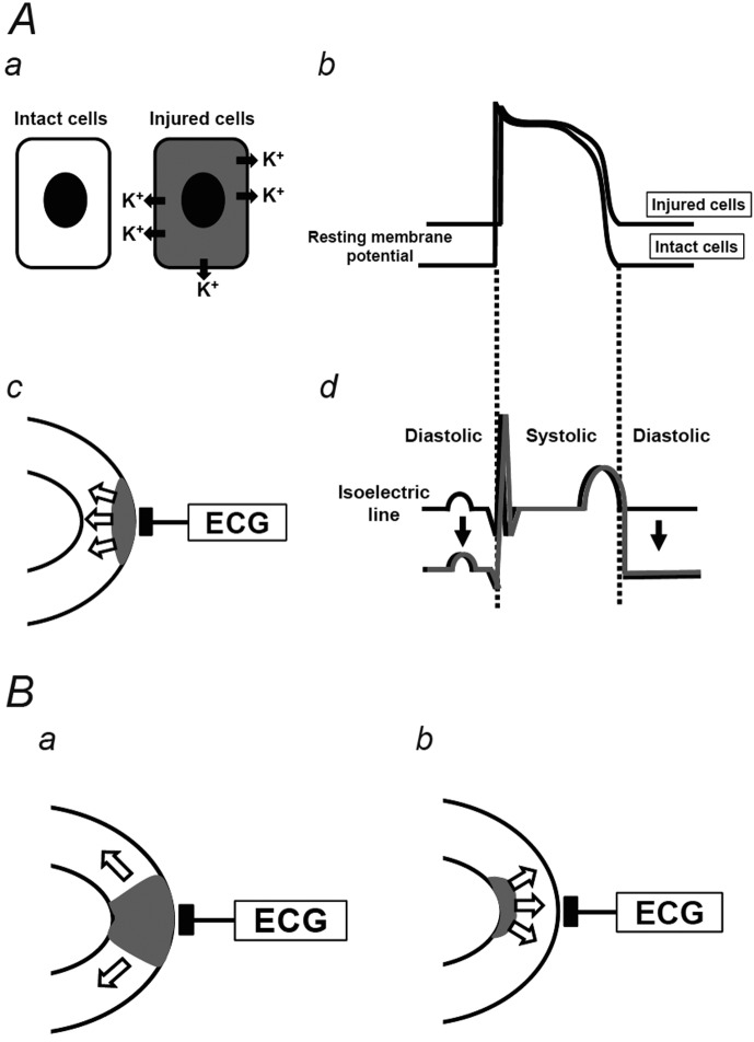
To mimic ischemic heart disease in humans, several animal models have been created, mainly in rodents by surgically ligating their coronary arteries. In the present study, by simply inducing burn injuries on the bullfrog heart, we reproduced abnormal ST segment changes in the electrocardiogram (ECG), mimicking those observed in ischemic heart disease, such as acute myocardial infarction and angina pectoris. The "currents of injury" created by a voltage gradient between the intact and damaged areas of the myocardium, negatively deflected the ECG vector during the diastolic phase, making the ST segment appear elevated during the systolic phase. This frog model of heart injury would be suitable to explain the mechanisms of ST segment changes observed in ischemic heart disease.
Figures


Similar articles
-
Subepicardial burn injuries in bullfrog heart induce electrocardiogram changes mimicking inferior wall myocardial infarction.J Vet Med Sci. 2022 Sep 1;84(9):1205-1210. doi: 10.1292/jvms.22-0243. Epub 2022 Jul 8. J Vet Med Sci. 2022. PMID: 35811132 Free PMC article.
-
Reciprocal ST segment changes reproduced in burn-induced subepicardial injury model in bullfrog heart.J Vet Med Sci. 2020 Feb 4;82(2):143-147. doi: 10.1292/jvms.19-0597. Epub 2019 Dec 10. J Vet Med Sci. 2020. PMID: 31827015 Free PMC article.
-
Partial exposure of frog heart to high-potassium solution: an easily reproducible model mimicking ST segment changes.J Vet Med Sci. 2018 Apr 18;80(4):578-582. doi: 10.1292/jvms.18-0010. Epub 2018 Mar 5. J Vet Med Sci. 2018. PMID: 29503350 Free PMC article.
-
Electorcardiographic ST-segment analysis in the characterization of myocardial ischemia and infarction.Circulation. 1976 Mar;53(3 Suppl):I73-81. Circulation. 1976. PMID: 767019 Review.
-
Electrocardiographic evaluation of reperfusion therapy in patients with acute myocardial infarction.Dan Med Bull. 1996 Feb;43(1):68-85. Dan Med Bull. 1996. PMID: 8906982 Review.
Cited by
-
Subepicardial burn injuries in bullfrog heart induce electrocardiogram changes mimicking inferior wall myocardial infarction.J Vet Med Sci. 2022 Sep 1;84(9):1205-1210. doi: 10.1292/jvms.22-0243. Epub 2022 Jul 8. J Vet Med Sci. 2022. PMID: 35811132 Free PMC article.
-
Insulin accelerates recovery from QRS complex widening in a frog heart model of hyperkalemia.J Vet Med Sci. 2021 Dec 2;83(12):1855-1859. doi: 10.1292/jvms.21-0481. Epub 2021 Oct 18. J Vet Med Sci. 2021. PMID: 34657900 Free PMC article.
-
Tree shrew (Tupaia belangeri) as a novel laboratory disease animal model.Zool Res. 2017 May 18;38(3):127-137. doi: 10.24272/j.issn.2095-8137.2017.033. Zool Res. 2017. PMID: 28585436 Free PMC article. Review.
-
Reciprocal ST segment changes reproduced in burn-induced subepicardial injury model in bullfrog heart.J Vet Med Sci. 2020 Feb 4;82(2):143-147. doi: 10.1292/jvms.19-0597. Epub 2019 Dec 10. J Vet Med Sci. 2020. PMID: 31827015 Free PMC article.
-
High-calcium exposure to frog heart: a simple model representing hypercalcemia-induced ECG abnormalities.J Vet Med Sci. 2017 Jan 20;79(1):71-75. doi: 10.1292/jvms.16-0413. Epub 2016 Oct 22. J Vet Med Sci. 2017. PMID: 27773880 Free PMC article.
References
-
- Fasciano R. W., 2nd, Tung L.1999. Factors governing mechanical stimulation in frog hearts. Am. J. Physiol. 277: H2311–H2320. - PubMed
MeSH terms
LinkOut - more resources
Full Text Sources
Other Literature Sources
Medical