. 2015 Sep 7:15:314.
doi: 10.1186/s12906-015-0826-x.

The anti-inflammatory and antifibrotic effects of Coreopsis tinctoria Nutt on high-glucose-fat diet and streptozotocin-induced diabetic renal damage in rats

Affiliations

The anti-inflammatory and antifibrotic effects of Coreopsis tinctoria Nutt on high-glucose-fat diet and streptozotocin-induced diabetic renal damage in rats

Lan Yao et al. BMC Complement Altern Med. .

Abstract

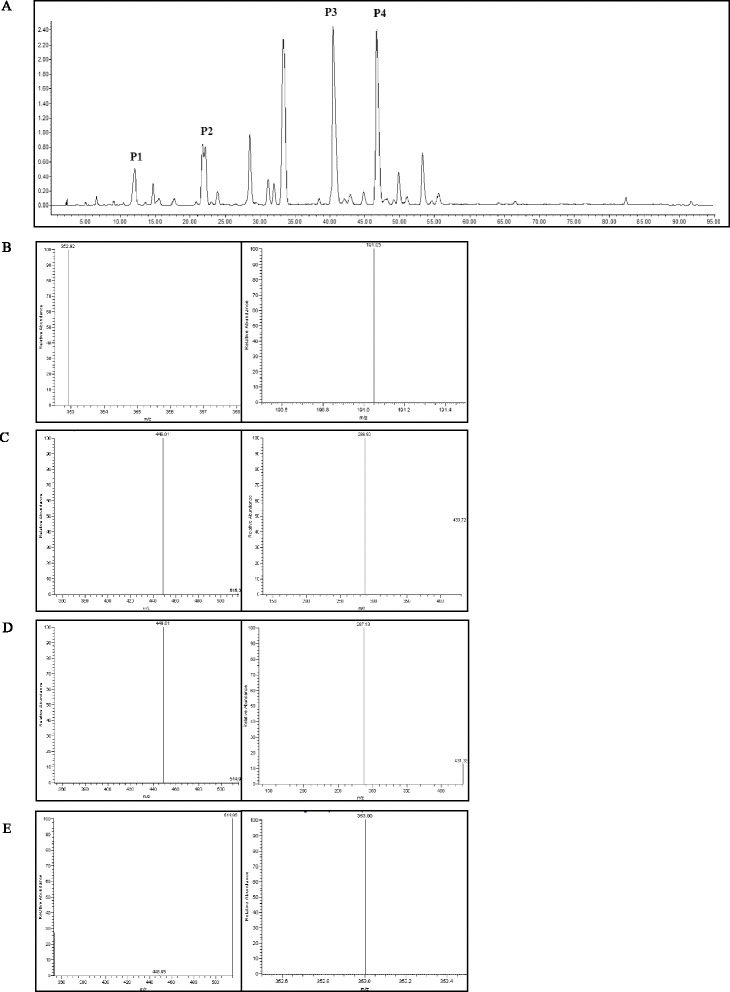
Background: Diabetic nephropathy is a serious complication of diabetes whose development process is associated with inflammation, renal hypertrophy, and fibrosis. Coreopsis tinctoria Nutt, traditionally used as a healthcare tea, has anti-inflammatory, anti-hyperlipidemia, and glycemic regulation activities. The aim of our study was to investigate the renal protective effect of ethyl acetate extract of C. tinctoria Nutt (AC) on high-glucose-fat diet and streptozotocin (STZ)-induced diabetic rats.

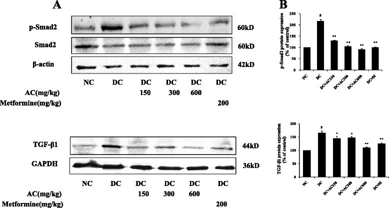
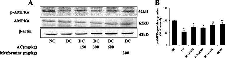
Methods: A diabetic rat model was induced by high-glucose-fat diet and intraperitoneal injection of 35 mg/kg STZ. After treatment with AC at a daily dose of 150, 300 or, 600 mg/kg for 4 weeks, metabolic and renal function parameters of serum and urine were examined. Degree of renal damage, renal proinflammatory cytokines, and fibrotic protein expression were analyzed by histopathology and immunohistochemistry. Renal AMP-activated protein kinase (AMPK) and transforming growth factor (TGF)-β1/Smad signaling pathway were determined by western blotting.

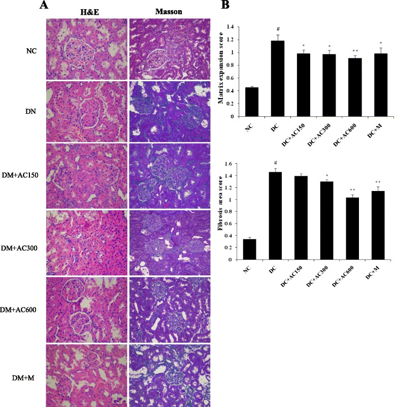
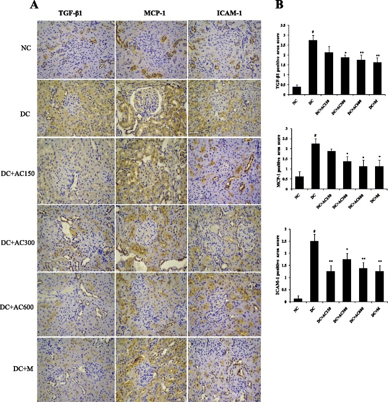
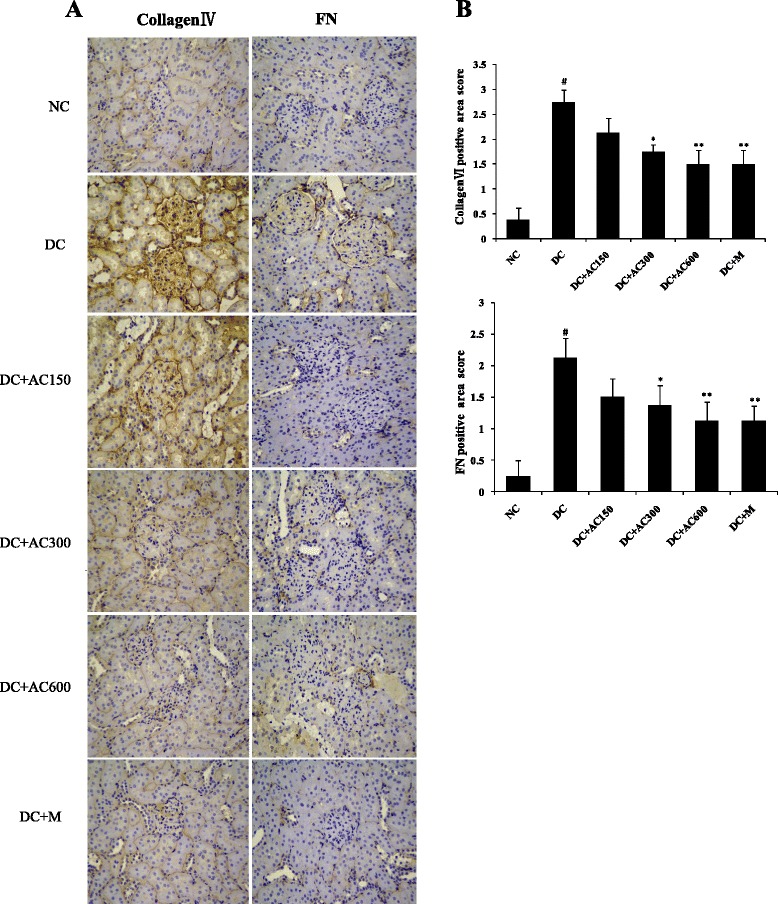
Results: Diabetic rats showed obvious renal dysfunction, inflammation and fibrosis. However, AC significantly reduced levels of blood glucose, total cholesterol, triglyceride, blood urea nitrogen, serum creatinine and urinary albumin, as well as expression of kidney proinflammatory cytokines of monocyte chemoattractant protein-1 and intercellular adhesion molecule-1. AC also ameliorated renal hypertrophy and fibrosis by reducing fibronectin and collagen IV and suppressing the TGF-β1/Smad signaling pathway. Meanwhile, AMPKα as a protective cytokine was markedly stimulated by AC.

Conclusion: In summary, AC controls blood glucose, inhibits inflammatory and fibrotic processes, suppresses the TGF-β1/Smad signaling pathway, and activates phosphorylation of AMPKα in the kidneys, which confirms the protective effects of AC in the early stage of diabetic kidney disease.


Figures

Fig. 1
Fig. 1
HPLC/MS analysis of AC. a HPLC analysis of AC at 280 nm wavelength. Four main compounds were (P1) chlorogenic acid, (P2) flavanomarein, (P3) marein, and (P4) 4,5-dicaffeoyl-quinic acid. Precursor ion (left) and product ion (right) of (b) chlorogenic acid, (c) flavanomarein, (d) marein, and (e) 4,5-dicaffeoyl-quinic acid
Fig. 2
Fig. 2
Renal histology after treatment with or without AC and metformin. a Pathological features of the left kidney tissue stained with H&E and Masson stain (orginal magnification 400×). Each photomicrograph represented the pathological feature of four rats from each group. b Matrix expansion and area of fibrosis among the groups were semiquantitatively analyzed. Values were expressed as mean ± SEM (n = 4). #P < 0.01 versus normal group; *P < 0.05 versus control group; **P < 0.01 versus control group
Fig. 3
Fig. 3
Expression of TGF-β1, MCP-1 and ICAM-1 in kidney. a Immunohistochemistry of TGF-β1, MCP-1 and ICAM-1 (orginal magnification 400×). b TGF-β1, MCP-1 and ICAM-1 expression was semiquantitatively analyzed. Values were expressed as mean ± SEM (n = 4). #P < 0.01 versus normal group; *P < 0.05 versus control group; **P < 0.01 versus control group
Fig. 4
Fig. 4
Expression of collagen IV and fibronectin in kidney. a Immunohistochemistry of collagen IV and fibronectin (orginal magnification 400×). b Collagen IV and fibronectin expression was semiquantitatively analyzed. Values were expressed as mean ± SEM (n = 4). #P < 0.01 versus normal group; *P < 0.05 versus control group; **P < 0.01 versus control group
Fig. 5
Fig. 5
Effect of AC on protein expression of TGF-β1, Smad2 and p-Smad2. a TGF-β1, Smad2 and p-Smad2 expression in right kidney. b Band density analysis of TGF-β1, Smad2 and p-Smad2. Values were expressed as mean ± SEM (n = 4). #P < 0.01 versus normal group; *P < 0.05 versus control group; **P < 0.01 versus control group
Fig. 6
Fig. 6
Effect of AC on protein expression of AMPK and p-AMPK. a AMPKand p-AMPK levels in right kidney. b Band density analysis of TGF-β1, AMPK and p-AMPK. Values were expressed as mean ± SEM (n = 3). #P < 0.01 versus normal group; *P < 0.05 versus control group; **P < 0.01 versus control group

References

    1. Chen X, Yang W. Epidemic trend of diabetes in China: For the Xiaoren Pan Distinguished Research Award in AASD. J Diabetes Investig. 2014;5:478–481. doi: 10.1111/jdi.12254. - DOI - PMC - PubMed
    1. Calcutt NA, Cooper ME, Kern TS, Schmidt AM. Therapies for hyperglycaemia-induced diabetic complications: from animal models to clinical trials. Nat Rev Drug Discov. 2009;8:417–429. doi: 10.1038/nrd2476. - DOI - PMC - PubMed
    1. Kanasaki K, Taduri G, Koya D. Diabetic nephropathy: the role of inflammation in fibroblast activation and kidney fibrosis. Front Endocrinol (Lausanne) 2013;4:7. - PMC - PubMed
    1. Soetikno V, Arozal W, Louisa M, Setiabudy R. New insight into the molecular drug target of diabetic nephropathy. Int J Endocrinol. 2014;2014:968681. doi: 10.1155/2014/968681. - DOI - PMC - PubMed
    1. Lim A. Diabetic nephropathy–complications and treatment. Int J Nephrol Renovasc Dis. 2014;7:361–381. doi: 10.2147/IJNRD.S40172. - DOI - PMC - PubMed

Publication types