Review
. 2015:2015:689404.
doi: 10.1155/2015/689404. Epub 2015 Aug 4.

Fractalkine Signaling and Microglia Functions in the Developing Brain

Affiliations
Review

Fractalkine Signaling and Microglia Functions in the Developing Brain

Isabelle Arnoux et al. Neural Plast. 2015.

Abstract

Microglial cells are the resident macrophages of the central nervous system (CNS). Besides their classical roles in pathological conditions, these immune cells also dynamically interact with neurons and influence their structure and function in physiological conditions. The neuronal chemokine fractalkine and its microglial receptor CX3CR1 are one important signaling pathway involved in these reciprocal interactions. In the present review, we will discuss recent evidence indicating that fractalkine signaling also determines several functions of microglial cells during normal CNS development. It has been known for a decade that microglial cells influence the neuronal death that normally occurs during CNS development. Surprisingly, recent evidence indicates that they can also support survival of developing neurons, control axon outgrowth, and laminar positioning of subsets of interneurons in the forebrain. Moreover, microglial cells influence the maturation of synaptic circuits at early postnatal stages: their phagocytic activity allows them to eliminate inappropriate synapses and they can also influence the functional expression of synaptic proteins by releasing mediators. Fractalkine signaling controls these functions of microglial cells in part by regulating their timely recruitment at sites of developing synapses. Finally, on-going research suggests that this signaling pathway is also a key player in neurodevelopmental disorders.


Figures

Figure 1
Figure 1
Fractalkine/CX3CR1 signaling controls survival of cortical neuron during early postnatal development. (a) Apoptotic cells revealed by TUNEL staining in the cortex of Cx3cr1 +/GFP (top) and Cx3cr1 GFP/GFP (bottom) mice at P5. Scale bar represents 100 μm. The arrowheads indicate TUNEL-positive elements in the layer V of the cortex. (b) Quantification of the TUNEL-positive cells density in different cortical layers. Note the increase of apoptotic cells in the layer V and II–IV in the cortex of Cx3cr1 GFP/GFP. Adapted from [43].
Figure 2
Figure 2
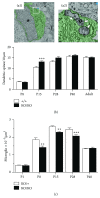
Fractalkine/CX3CR1 signaling modulates synaptic pruning by microglia during postnatal development. (a) Microglial cells remove presynaptic elements by synaptic pruning at P5 in the retinogeniculate system. (a1) Low magnification electronic microscopy of microglia. Asterisks denote the nucleus and the cytoplasm is pseudocolored green. Scale bar = 1 μm. (a2) Magnified regions of (a1) (white box) demonstrating membrane-bound elements engulfed by microglia. Arrows designate elements containing presynaptic machinery (40 nm vesicles). The arrowhead designates engulfed material resembling juxtaposed postsynaptic elements. Scale bar = 100 nm. Adapted from [53]. (b) A transient increase in dendritic spine density was observed in CX3CR1 deficient (KO/KO) mice when compared with WT (+/+) littermates during the second postnatal week (∗∗∗ p < 0.0001). This transient increase in dendritic spines number could result of a transient deficit of synaptic pruning. (c) Quantification of microglia nuclei in the CA1 stratum radiatum from the hippocampus revealed a transient decrease in microglia density in CX3CR1 deficient mice at P8, P15, and P28 compared with control littermates (∗∗ p < 0.005). This decrease in microglia number in KO mice suggests a transient delayed microglia recruitment which can explain the transient deficit of synaptic pruning. ((b) and (c)) Adapted from [47].
Figure 3
Figure 3
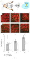
Fractalkine/CX3CR1 signaling controls the recruitment of microglia and the functional maturation of thalamocortical synapses. (a) Drawing of the sensory system of vibrissae in rodents and link between the distribution of vibrissae and that of barrels in layer 4 somatosensory cortex. Adapted from [59]. ((b) and (c)) Microglia (green) distribution in the layer 4 of the somatosensory cortex during postnatal development in Cx3cr1 +/eGFP (b) and Cx3cr1 eGFP/eGFP (c) mice. At P5, microglial cells are exclusively located outside of the barrel centers which contain thalamocortical synapses (red, staining of thalamocortical axons). At P7, microglial cells start to invade the barrel centers in Cx3cr1 +/eGFP mice and this invasion is delayed in Cx3cr1 eGFP/eGFP mice. At P9, microglial cell distribution is similar for the two genotypes. Scale bar, 100 µm. (d) Relative change in the synaptic currents resulting of the activation of AMPA (α-amino-3-hydroxy-5-methyl-4-isoxazolepropionic acid) and NMDA (N-methyl-D-aspartate) postsynaptic receptors expressed at thalamocortical synapses between P5 and P9 in Cx3cr1 +/eGFP and Cx3cr1 eGFP/eGFP mice. Note that the AMPA/NMDA ratio increases between P5 and P9 in Cx3cr1 +/eGFP but not in Cx3cr1 eGFP/eGFP mice. ((b) and (c)) Adapted from [48].
Figure 4
Figure 4
Impaired microglia motility in developing CX3CR1 deficient mice. (a) Two-photon images in an acute slice of a P7 Cx3cr1 +/eGFP mouse showing the dynamics of microglial processes and soma after the insertion (at time 0 min, not shown) of a pipette (red dot) containing 2-MeSADP (100 µM). Yellow arrowheads indicate the soma of 2 microglial cells moving toward the pipette. Green arrow indicates a retracting process. Calibration bar is 10 µm. (b) Comparison of the mean velocity of microglia soma toward the 2-MeSADP-containing pipette for Cx3cr1 +/eGFP (50 cells, 6 experiments) Cx3cr1 +/eGFP (42 cells, 5 experiments) animals ( p < 0.05, ∗∗ p < 0.01, Mann-Whitney test).

References

    1. Tremblay M.-È., Stevens B., Sierra A., Wake H., Bessis A., Nimmerjahn A. The role of microglia in the healthy brain. The Journal of Neuroscience. 2011;31(45):16064–16069. doi: 10.1523/jneurosci.4158-11.2011. - DOI - PMC - PubMed
    1. Kettenmann H., Kirchhoff F., Verkhratsky A. Microglia: new roles for the synaptic stripper. Neuron. 2013;77(1):10–18. doi: 10.1016/j.neuron.2012.12.023. - DOI - PubMed
    1. Salter M. W., Beggs S. Sublime microglia: expanding roles for the guardians of the CNS. Cell. 2014;158:15–24. doi: 10.1016/j.cell.2014.06.008. - DOI - PubMed
    1. Kierdorf K., Erny D., Goldmann T., et al. Microglia emerge from erythromyeloid precursors via Pu.1-and Irf8-dependent pathways. Nature Neuroscience. 2013;16(3):273–280. doi: 10.1038/nn.3318. - DOI - PubMed
    1. Ginhoux F., Greter M., Leboeuf M., et al. Fate mapping analysis reveals that adult microglia derive from primitive macrophages. Science. 2010;330(6005):841–845. doi: 10.1126/science.1194637. - DOI - PMC - PubMed

Publication types

Substances

LinkOut - more resources