Review
. 2015 Aug 19:6:230.
doi: 10.3389/fphys.2015.00230. eCollection 2015.

Stress, glucocorticoid hormones, and hippocampal neural progenitor cells: implications to mood disorders

Affiliations
Review

Stress, glucocorticoid hormones, and hippocampal neural progenitor cells: implications to mood disorders

Tomoshige Kino. Front Physiol. .

Abstract

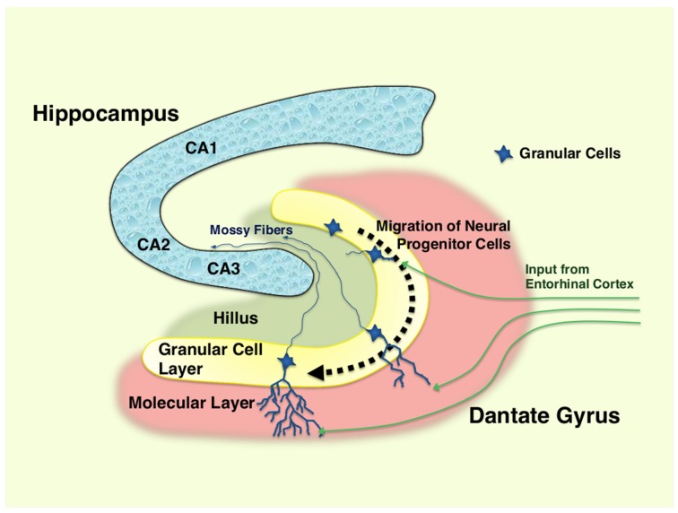
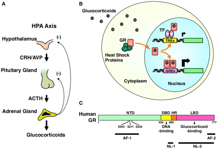
The hypothalamic-pituitary-adrenal (HPA) axis and its end-effectors glucocorticoid hormones play central roles in the adaptive response to numerous stressors that can be either internal or external. Thus, this system has a strong impact on the brain hippocampus and its major functions, such as cognition, memory as well as behavior, and mood. The hippocampal area of the adult brain contains neural stem cells or more committed neural progenitor cells, which retain throughout the human life the ability of self-renewal and to differentiate into multiple neural cell lineages, such as neurons, astrocytes, and oligodendrocytes. Importantly, these characteristic cells contribute significantly to the above-indicated functions of the hippocampus, while various stressors and glucocorticoids influence proliferation, differentiation, and fate of these cells. This review offers an overview of the current understanding on the interactions between the HPA axis/glucocorticoid stress-responsive system and hippocampal neural progenitor cells by focusing on the actions of glucocorticoids. Also addressed is a further discussion on the implications of such interactions to the pathophysiology of mood disorders.

Keywords: dentate gyrus; glucocorticoid receptor; hippocampus; major depression; neural stem cells (NSCs).


Figures

Figure 1
Figure 1
Hippocampus and differentiation of neural progenitor cells in dentate gyrus. Neural progenitor cells reside in the granular layer (subgranular zone) of the dentate gyrus as a mixture with immature and mature granular cell neurons. Following the differentiation (~1–2 months), immature neurons generated from neural progenitor cells migrate into the lower arm of the dentate gyrus, and extend their dendrites into the molecular layer, which receives excitatory neurons from the entorhinal cortex. They also spread into the hillus their excitatory mossy fibers, and form synaptic connections to the pyramidal neurons in the CA3 region of the hippocampus. Over half of the immature neurons die during their differentiation process. CA1, 2, and 3: region 1, 2, and 3 of the hippocampal proper.
Figure 2
Figure 2
HPA axis and GR. (A) Organization of the HPA axis. The HPA axis consists of three components, PVN of the hypothalamus, the anterior pituitary gland, and the adrenal cortex. Neurons residing in PVN produce CRH and AVP, and release them into the pituitary portal system under the control of the upper regulatory centers, such as the central circadian rhythm center hypothalamic suprachiasmatic nucleus (SCN) and the stress responsive hippocampus, prefrontal cortex, and the amygdala. CRH and AVP stimulate the secretion of ACTH from the corticotrophs of the anterior pituitary gland. Circulating ACTH then stimulates the production and the secretion of glucocorticoids (cortisol in humans and corticosterone in rodents) from the adrenocortical cells located in the zona fasciculata of the adrenal gland. Secreted glucocorticoids then suppress the upper regulatory centers including PVN and the pituitary gland, forming a closed regulatory loop. (B) Intracellular circulation of the GR. In the absence of glucocorticoids, GR resides in the cytoplasm forming a heterocomplex with several heat shock proteins (HSP), including HSP90, 70, and 23. Upon binding to glucocorticoids, GR releases HSPs, exposes its nuclear localization signals (NLs) to the nuclear pore complex and translocates into the nucleus. In the nucleus, GR directly binds as a homodimer its specific recognition sequences called glucocorticoid response elements (GREs), which are located in the promoter region of glucocorticoid-responsive genes, and stimulates their transcriptional activity by attracting many transcriptional cofactors and the RNA polymerase II complex. GR also modulates the transcriptional activity of other transcription factors through physical protein-protein interactions without associating directly to DNA. After regulating the transcription of glucocorticoid-responsive genes, GR moves back into the cytoplasm with the help of the nuclear export system and returns to its ligand friendly condition by reforming a heterocomplex with HSPs. (C) Linearized protein structure of the human GR and its functional distribution. Human GR consists of 777 amino acids and composes of three subdomains, the N-terminal or immunogenic domain (NTD), middle DNA-binding domain (DBD), and the C-terminal ligand-binding domain (LBD). Between DBD and LBD, there is a small area called the hinge region (HR). GR has two transactivation domains, activation function (AF)-1 and -2, which are respectively located in NTD and LBD. GR also has two nuclear localization signals (NL)-1 and -2. NL-1 is located in the DBD-HR boarder and mediates the rapid nuclear translocation of GR by communicating with the importin α/β nuclear pore complex, while NL-2 distributes in the entire LBD and mediates slow nuclear translocation of this receptor. GR has three serine residues (serines 203, 211, and 226) in the AF-1 of NTD, which are phosphorylated by several serine/threonine-directed protein kinases including CDK5. ACTH, adrenocorticotropic hormone; AF-1 and -2, activation function-1 and -2; AVP, arginine vasopressin; CRH, corticotropin-releasing hormone; DBD, DNA-binding domain; GR, glucocorticoid receptor; GREs, glucocorticoid response elements; HPA axis, hypothalamic-pituitary-adrenal axis; HR, hinge region; HSPs, heat shock proteins; LBD, ligand-binding domain; NL-1 and -2, nuclear localization signal-1 and -2; NTD, N-terminal domain; S203, 211, and 226, serine at amino acid position 203, 211, and 226; TF, transcription factor; TREs, transcription factor response elements.

References

    1. Anacker C., Zunszain P. A., Cattaneo A., Carvalho L. A., Garabedian M. J., Thuret S., et al. . (2011). Antidepressants increase human hippocampal neurogenesis by activating the glucocorticoid receptor. Mol. Psychiatry 16, 738–750. 10.1038/mp.2011.26 - DOI - PMC - PubMed
    1. Androutsellis-Theotokis A., Chrousos G. P., McKay R. D., DeCherney A. H., Kino T. (2013). Expression profiles of the nuclear receptors and their transcriptional coregulators during differentiation of neural stem cells. Horm. Metab. Res. 45, 159–168. 10.1055/s-0032-1321789 - DOI - PMC - PubMed
    1. Androutsellis-Theotokis A., Murase S., Boyd J. D., Park D. M., Hoeppner D. J., Ravin R., et al. . (2008a). Generating neurons from stem cells. Methods Mol. Biol. 438, 31–38. 10.1007/978-1-59745-133-8_4 - DOI - PubMed
    1. Androutsellis-Theotokis A., Rubin de Celis M. F., Ehrhart-Bornstein M., Bornstein S. R. (2012). Common features between chromaffin and neural progenitor cells. Mol. Psychiatry 17, 351. 10.1038/mp.2012.18 - DOI - PubMed
    1. Androutsellis-Theotokis A., Rueger M. A., Mkhikian H., Korb E., McKay R. D. (2008b). Signaling pathways controlling neural stem cells slow progressive brain disease. Cold Spring Harb. Symp. Quant. Biol. 73, 403–410. 10.1101/sqb.2008.73.018 - DOI - PubMed

LinkOut - more resources