. 2015 Aug 18:6:177.
doi: 10.3389/fneur.2015.00177. eCollection 2015.

A Prospective Pilot Trial for Pallidal Deep Brain Stimulation in Huntington's Disease

Affiliations

A Prospective Pilot Trial for Pallidal Deep Brain Stimulation in Huntington's Disease

Lars Wojtecki et al. Front Neurol. .

Abstract

Background: Movement disorders in Huntington's disease are often medically refractive. The aim of the trial was assessment of procedure safety of deep brain stimulation, equality of internal- and external-pallidal stimulation and efficacy followed-up for 6 months in a prospective pilot trial.

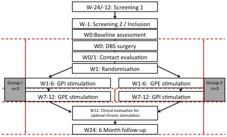
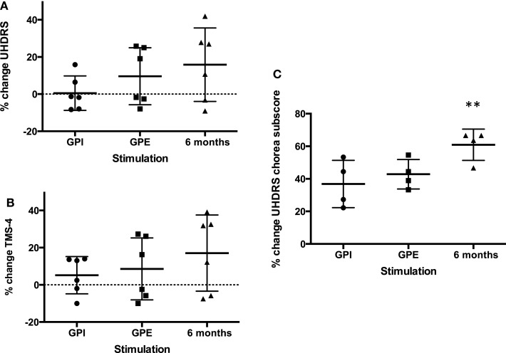
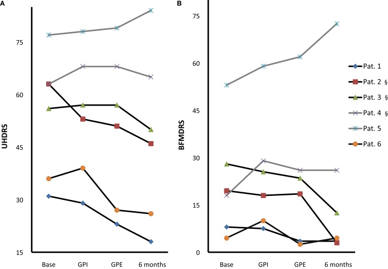
Methods: In a controlled double-blind phase six patients (four chorea-dominant, two Westphal-variant) with predominant movement disorder were randomly assigned to either the sequence of 6-week internal- or 6-week external-pallidal stimulation, or vice versa, followed by further 3 months chronic pallidal stimulation at the target with best effect-side-effect ratio. Primary endpoints were changes in the Unified Huntington's Disease Rating Scale motor-score, chorea subscore, and total motor-score 4 (blinded-video ratings), comparing internal- versus external-pallidal stimulation, and 6 months versus baseline. Secondary endpoints assessed scores on dystonia, hypokinesia, cognition, mood, functionality/disability, and quality-of-life.

Results: Intention-to-treat analysis of all patients (n = 3 in each treatment sequence): Both targets were equal in terms of efficacy. Chorea subscores decreased significantly over 6 months (-5.3 (60.2%), p = 0.037). Effects on dystonia were not significant over the group due to it consisting of three responders (>50% improvement) and three non-responders. Westphal patients did not improve. Cognition was stable. Mood and some functionality/disability and quality-of-life scores improved significantly. Eight adverse events and two additional serious adverse events - mostly internal-pallidal stimulation-related - resolved without sequalae. No procedure-related complications occurred.

Conclusion: Pallidal deep brain stimulation was demonstrated to be a safe treatment option for the reduction of chorea in Huntington's disease. Their effects on chorea and dystonia and on quality-of-life should be examined in larger controlled trials.

Keywords: chorea; deep brain stimulation; pallidum; Huntington's Disease.


Figures

Figure 1
Figure 1
Flow chart of the study. n = 6, W: week, M: month, dashed red lines illustrate timepoints of study assessments, red arrows illustrate calculated comparisons.
Figure 2
Figure 2
Percent change improvement from baseline of primary endpoints. Mean with SD (error bars) and individual results (A): UHDRS, (B): TMS-4, (C): UHDRS chorea subscore, note: chorea subscore could only be calculated for four patients (non-Westphal). **Significant change from baseline, p < 0.01.
Figure 3
Figure 3
Individual motor score changes. Individual UHDRS (A) and BFMDRS (B) at baseline, with GPI, GPE and chronic 6-month stimulation [Pat. #1, 5, 6 GPE; Pat. #2, 3, 4 GPI (§)]; Westphal patients: gray lines, #4, 5. Note: GPI/GPE sequence was randomized with GPI first in Pat. #1, 4, 5 and GPE first in Pat. #2, 3, 6.
Figure 4
Figure 4
Mean electrode localization. Visualization of mean coordinates of left and right hemisphere mirrored to the left; 3D space relative to AC-PC line (green dot: AC, red dot: PC), gray mash: GPI, green mash: GPE; lowermost contacts comprise GPI and uppermost contacts comprise GPE stimulation. Thus, mean chronic stimulation at 6-month follow-up projects mid-electrode to the border zone between GPE and GPI. For visualization the following atlas software was used: Medtronic DBS Neurosurgical Simulator, licensed 2008, Version 1.2.3, Medtronic Inc., Minneapolis, MN, USA.
Figure 5
Figure 5
Individual electrode localization. Visualization on 3D coronary MRI-view of individual electrodes and volume of tissue activated (VTA, in red) in relation to the pallidum (in brown) for the ventral target (GPI), the dorsal target (GPE) and the target at 6-month follow-up.

References

    1. Krack P, Pollak P, Limousin P, Hoffmann D, Benazzouz A, Le Bas JF, et al. Opposite motor effects of pallidal stimulation in Parkinson’s disease. Ann Neurol (1998) 43:180–92.10.1002/ana.410430208 - DOI - PubMed
    1. Kupsch A, Benecke R, Muller J, Trottenberg T, Schneider GH, Poewe W, et al. Pallidal deep-brain stimulation in primary generalized or segmental dystonia. N Engl J Med (2006) 355:1978–90.10.1056/NEJMoa063618 - DOI - PubMed
    1. Edwards TC, Zrinzo L, Limousin P, Foltynie T. Deep brain stimulation in the treatment of chorea. Mov Disord (2012) 27:357–63.10.1002/mds.23967 - DOI - PubMed
    1. López-Sendón Moreno JL, García-Caldentey J, Regidor I, Alamo MD, García de Yébenes J. A 5-year follow-up of deep brain stimulation in Huntington’s disease. Parkinsonism Relat Disord (2014) 20:260–1.10.1016/j.parkreldis.2013.11.007 - DOI - PubMed
    1. Spielberger S, Hotter A, Wolf E, Eisner W, Müller J, Poewe W, et al. Deep brain stimulation in Huntington’s disease: a 4-year follow-up case report. Mov Disord (2012) 27:806–7.10.1002/mds.24959 - DOI - PubMed

LinkOut - more resources