Review
. 2015 Aug 18:6:422.
doi: 10.3389/fimmu.2015.00422. eCollection 2015.

DAMPs from Cell Death to New Life

Affiliations
Review

DAMPs from Cell Death to New Life

Emilie Vénéreau et al. Front Immunol. .

Abstract

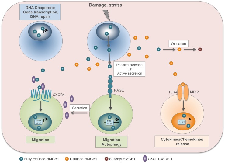
Our body handles tissue damage by activating the immune system in response to intracellular molecules released by injured tissues [damage-associated molecular patterns (DAMPs)], in a similar way as it detects molecular motifs conserved in pathogens (pathogen-associated molecular patterns). DAMPs are molecules that have a physiological role inside the cell, but acquire additional functions when they are exposed to the extracellular environment: they alert the body about danger, stimulate an inflammatory response, and finally promote the regeneration process. Beside their passive release by dead cells, some DAMPs can be secreted or exposed by living cells undergoing a life-threatening stress. DAMPs have been linked to inflammation and related disorders: hence, inhibition of DAMP-mediated inflammatory responses is a promising strategy to improve the clinical management of infection- and injury-elicited inflammatory diseases. However, it is important to consider that DAMPs are not only danger signals but also central players in tissue repair. Indeed, some DAMPs have been studied for their role in tissue healing after sterile or infection-associated inflammation. This review is focused on two exemplary DAMPs, HMGB1 and adenosine triphosphate, and their contribution to both inflammation and tissue repair.

Keywords: ATP; DAMP; HMGB1; inflammation; tissue repair.


Figures

Figure 1
Figure 1
HMGB1 is a redox-sensitive DAMP. In the nucleus, fully reduced HMGB1 acts as a DNA chaperone and contributes to gene transcription and DNA repair. Upon injury or stress, HMGB1 is passively released by dead cells or actively secreted by stressed cells. The fully reduced HMGB1 binds to CXCL12 chemokine to form a heterocomplex, which in turn binds to CXCR4 and induces cell migration. In addition, HMGB1 interacts with RAGE to induce CXCL12 secretion and autophagy. In the extracellular compartment, disulfide HMGB1 derives from the active secretion and/or the conversion of fully reduced HMGB1 by oxidation. Disulfide HMGB1 binds to TLR4/MD-2 complex and induces cytokine/chemokine release. Finally, HMGB1 cysteines are terminally oxidized to sulfonates; sulfonyl-HMGB1 is neither chemoattractant nor has cytokine-inducing activity.
Figure 2
Figure 2
ATP is a time-resolved DAMP. In the cell, ATP derived from mitochondria is a universal energy source in cell reactions and metabolism. Upon damage or stress, ATP, and other nucleotides, are passively released by dead cells or actively secreted by stressed cells. ATP binds to ionotropic P2X receptors (P2XR), which are nucleotide-gated ion channels, allowing sodium (Na+) and calcium (Ca2+) influx and potassium (K+) efflux. The increased level of intracellular calcium activates p38 MAPK or phospholipase A2 signaling, while potassium efflux activates the inflammasome. P2XR signaling is involved in inflammation, tumor and bacterial killing. ATP also binds to metabotropic P2Y receptors (P2YR), which are G-protein-coupled, and induces activation of MAPK and phospholipase C (PLC). P2YR signaling is implicated in inflammation and wound healing, and ATP released by apoptotic cells acts as a “find-me” signal to recruit macrophages to the site of damage and to promote clearance of apoptotic cells. ATP signaling is abolished by the enzymatic conversion of ATP to adenosine through the ecto-nucleoside triphosphate diphosphohydrolase CD39 (from ATP to AMP) and the ecto-5′-nucleotidase CD73 (from AMP to adenosine).
Figure 3
Figure 3
HMGB1 and ATP in tissue repair. Following tissue injury, HMGB1 and ATP are passively released by dead cells or actively secreted by stressed cells. Then, they recruit to the site of damage the cell types required to heal the wound. First, immune cells are needed to clean the wound by engulfing dead cells and cellular debris. Then, stem cells and neighboring cells are induced to proliferate and build new tissue, together with its extracellular matrix. Endothelial cells are activated to form new blood vessels.

Similar articles

Cited by

References

    1. Janeway CA. Approaching the asymptote? Evolution and revolution in immunology. Cold Spring Harb Symp Quant Biol (1989) 54(Pt 1):1–13.10.1101/SQB.1989.054.01.003 - DOI - PubMed
    1. Matzinger P. Tolerance, danger, and the extended family. Annu Rev Immunol (1994) 12:991–1045.10.1146/annurev.iy.12.040194.005015 - DOI - PubMed
    1. Land W, Schneeberger H, Schleibner S, Illner WD, Abendroth D, Rutili G, et al. The beneficial effect of human recombinant superoxide dismutase on acute and chronic rejection events in recipients of cadaveric renal transplants. Transplantation (1994) 57:211–7.10.1097/00007890-199401001-00010 - DOI - PubMed
    1. Scaffidi P, Misteli T, Bianchi ME. Release of chromatin protein HMGB1 by necrotic cells triggers inflammation. Nature (2002) 418:191–5.10.1038/nature00858 - DOI - PubMed
    1. Shi Y, Evans JE, Rock KL. Molecular identification of a danger signal that alerts the immune system to dying cells. Nature (2003) 425:516–21.10.1038/nature01991 - DOI - PubMed