. 2015:2015:931782.
doi: 10.1155/2015/931782. Epub 2015 Aug 10.

Luteolin Inhibits Angiotensin II-Stimulated VSMC Proliferation and Migration through Downregulation of Akt Phosphorylation

Affiliations

Luteolin Inhibits Angiotensin II-Stimulated VSMC Proliferation and Migration through Downregulation of Akt Phosphorylation

Tongda Xu et al. Evid Based Complement Alternat Med. 2015.

Abstract

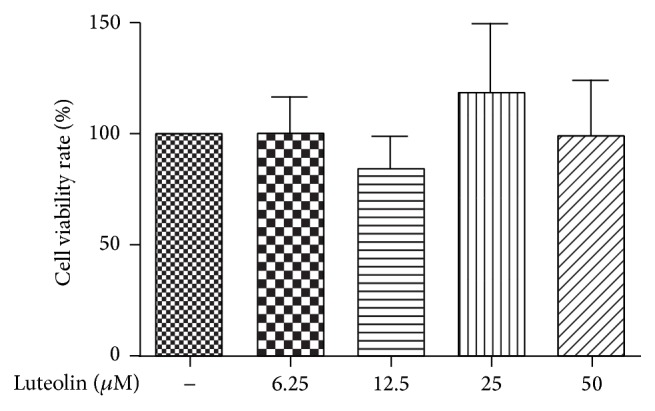
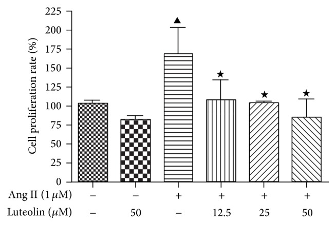
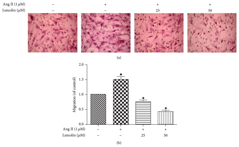
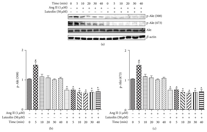
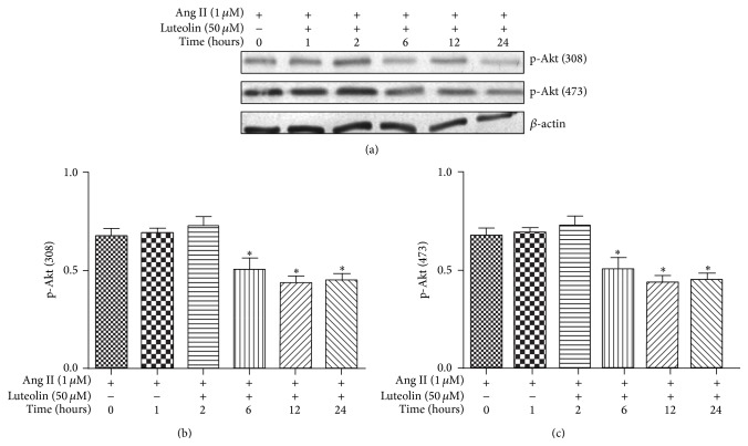
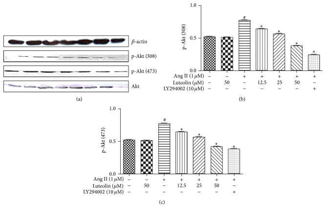
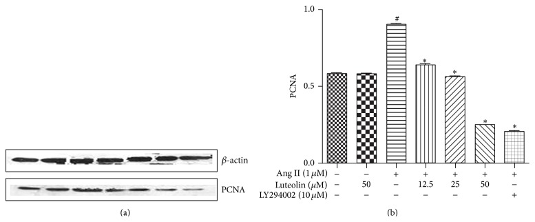
Luteolin is a naturally occurring flavonoid found in many plants that possesses cardioprotective properties. The purpose of this study was to elucidate the effect of luteolin on vascular smooth muscle cells (VSMCs) proliferation and migration induced by Angiotensin II (Ang II) and to investigate the mechanism(s) of action of this compound. Rat VSMCs were cultured in vitro, and the proliferation and migration of these cells following Ang II stimulation were monitored. Different doses of luteolin were added to VSMC cultures, and the proliferation and migration rate were observed by MTT and Transwell chamber assays, respectively. In addition, the expressions of p-Akt (308), p-Akt (473), and proliferative cell nuclear antigen (PCNA) in VSMCs were monitored by Western blotting. This study demonstrated that luteolin has an inhibitory effect on Ang II-induced VSMC proliferation and migration. Further, the levels of p-Akt (308), p-Akt (473), and PCNA were reduced in VSMCs treated with both Ang II and luteolin compared to VSMCs treated with only Ang II. These findings strongly suggest that luteolin inhibits Ang II-stimulated proliferation and migration of VSMCs, which is partially due to downregulation of the Akt signaling pathway.


Figures

Figure 1
Figure 1
Investigating the cytotoxicity of luteolin in VSMCs. VSMCs were incubated with varying concentrations of luteolin (6.25, 12.5, 25, and 50 μM) for 12 h, MTT reagent was added, and the OD value of each group was measured. The cell viability of the control group was taken as 100%.
Figure 2
Figure 2
Inhibitive effect of luteolin on VSMCs proliferation induced by Ang II. VSMCs were pretreated with the indicated concentrations of luteolin for 12 h prior to being stimulated with 1 μM Ang II. MTT assays were performed to measure cell proliferation, and the cell proliferation rate of the control group was taken as 100%. Data are shown as the mean ± SEM of 3 independent experiments. P < 0.05 compared with the control group; P < 0.05 compared with the Ang II group.
Figure 3
Figure 3
Inhibitory effect of luteolin on Ang II-induced VSMC migration. After being incubated with the indicated concentrations of luteolin, VSMCs were stimulated with 1 μM Ang II. The VSMC migration rate was determined by Transwell chamber assay. Bright-field images of randomly selected squares per group (×100). The cell migration rate of the control group was taken as 1. P < 0.05 compared with the control group; P < 0.05 compared with the Ang II group.
Figure 4
Figure 4
Effect of luteolin on different lengths of time on Ang II-induced activation of Akt. Following pretreatment with 50 μM luteolin for 12 h, VSMCs were stimulated with Ang II (1 μM) for the indicated times, and luteolin significantly attenuates Akt phosphorylation induced by Ang II. The cells were lysed and analyzed with antibodies against p-Akt (308), p-Akt (473), and Akt. # P < 0.05 compared with the control group; P < 0.05 compared with the Ang II group.
Figure 5
Figure 5
Effect of luteolin for different lengths of times on Ang II-induced activation of Akt. Following pretreatment with luteolin for different lengths of times, VSMCs were incubated with 1 μM Ang II for 12 h. The cells were lysed and analyzed with the antibodies against p-Akt (308), p-Akt (473), and Akt. Equal protein loading was confirmed by comparison of β-actin levels. P < 0.05 compared with the Ang II group.
Figure 6
Figure 6
Effect of different concentrations of luteolin on Ang II-induced activation of Akt. After incubation with the indicated concentrations of luteolin, VSMCs were stimulated with 1 μM Ang II. The cells were lysed and analyzed using the antibodies versus p-Akt (308), p-Akt (473), and Akt. Equal protein loading was confirmed by comparison of β-actin levels. # P < 0.05 compared with the control group; P < 0.05 compared with the Ang II group.
Figure 7
Figure 7
Inhibitory effects of luteolin on PCNA expression in Ang II-stimulated VSMCs. The cells were lysed and analyzed using the antibodies versus PCNA. Equal protein loading was confirmed by comparison of β-actin levels. # P < 0.05 compared with the control group; P < 0.05 compared with the Ang II group.

Similar articles

Cited by

References

    1. Libby P. Inflammation in atherosclerosis. Nature. 2002;420(6917):868–874. doi: 10.1038/nature01323. - DOI - PubMed
    1. Alvarez E., Rodiño-Janeiro B. K., Ucieda-Somoza R., González-Juanatey J. R. Pravastatin counteracts angiotensin II-induced upregulation and activation of nadph oxidase at plasma membrane of human endothelial cells. Journal of Cardiovascular Pharmacology. 2010;55(2):203–212. doi: 10.1097/FJC.0b013e3181ce5f5a. - DOI - PubMed
    1. Herr D., Rodewald M., Fraser H. M., et al. Regulation of endothelial proliferation by the renin-angiotensin system in human umbilical vein endothelial cells. Reproduction. 2008;136(1):125–130. doi: 10.1530/REP-07-0374. - DOI - PubMed
    1. Doran A. C., Meller N., McNamara C. A. Role of smooth muscle cells in the initiation and early progression of atherosclerosis. Arteriosclerosis, Thrombosis, and Vascular Biology. 2008;28(5):812–819. doi: 10.1161/atvbaha.107.159327. - DOI - PMC - PubMed
    1. Gorenne I., Kavurma M., Scott S., Bennett M. Vascular smooth muscle cell senescence in atherosclerosis. Cardiovascular Research. 2006;72(1):9–17. doi: 10.1016/j.cardiores.2006.06.004. - DOI - PubMed

LinkOut - more resources