. 2015:2015:375947.
doi: 10.1155/2015/375947. Epub 2015 Aug 12.

The Diagnostic and Therapeutic Challenges of Posttraumatic Iris Implantation Cysts: Illustrative Case Presentations and a Review of the Literature

Affiliations

The Diagnostic and Therapeutic Challenges of Posttraumatic Iris Implantation Cysts: Illustrative Case Presentations and a Review of the Literature

Nandini Venkateswaran et al. Case Rep Ophthalmol Med. 2015.

Abstract

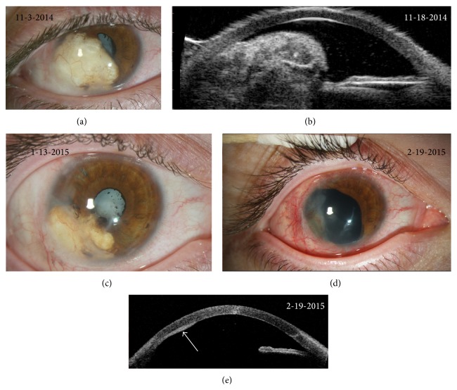
Posttraumatic iris implantation cysts are rare ocular findings that are often associated with poor visual outcomes. Iris implantation cysts can present clinicians with diagnostic and therapeutic challenges given their variable presentations and frequently destructive nature. In this paper, we provide descriptions of two unusual cases of posttraumatic iris implantation cysts. The first case is of a recurrent keratin-filled iris implantation cyst that developed after open globe injury and intraocular implantation of cilia and was treated with cyst debulking procedures, injections of 5-Fluorouracil, and iridocyclectomy. The second case is of recurrent posttraumatic serous iris implantation cysts that were treated with laser, cyst aspiration, and injections of 5-Fluorouracil. We use these cases as a platform to discuss the different manifestations of implantation cysts, the roles of anterior segment optical coherence tomography, ultrasound biomicroscopy, and histopathology in facilitating timely and accurate diagnosis and review the range of available therapeutic modalities. We discuss conservative treatment approaches, including the novel use of 5-Fluorouracil therapy as an adjunct therapy, as well as more aggressive surgical excision requiring ocular reconstruction. Through a discussion of these cases and review of the literature, we provide recommendations to assist clinicians in managing this uncommon but vision-threatening condition and minimizing complications.


Figures

Figure 1
Figure 1
Clinical and diagnostic images from case 1: initial presentation. (a) Slit-lamp photograph of the patient's right eye upon presentation two years after his initial penetrating ocular injury with a wire fence. An inferocentral corneal scar is seen (asterisk). The pupil is irregular with a large, opaque, iris cyst extending from 7 to 9 o'clock with two embedded cilia (arrows). The lens is clear with no signs of cataract formation. (b) Anterior segment optical coherence tomography (AS-OCT) images illustrating the healed corneal laceration (arrow) from the patient's prior penetrating ocular injury and the location and contours of the iris implantation cyst. The cyst abuts the posterior cornea and there is absence of normal angle architecture. High reflectivity within the cyst indicates that the lesion is unlikely to be fluid-filled. (c) Slit-lamp photograph of the patient's eye one week after debulking of the cyst and injection of 5-Fluorouracil (5-FU) within the cyst cavity. The cyst was approached through a temporal scleral tunnel incision to facilitate precise entry into the body of the cyst for drainage and prevent expulsion of debris into the anterior chamber. Following surgery, the cyst initially resolved and there is rounding of the pupil.
Figure 2
Figure 2
Clinical and diagnostic images from case 1: subsequent presentation. (a) Slit-lamp photograph illustrating recurrence of the iris cyst along the inferotemporal iris after the patient was lost to follow-up for 1.5 years. Posterior synechiae have developed and the pupil is largely obstructed by the iris cyst. A white cataract can be seen through the small pupillary opening. (b) Ultrasound biomicroscopic images demonstrating copious echogenic material within the iris cyst. Normal iris and angle structure are disrupted and the cyst abuts the ciliary body. (c) Slit-lamp photograph showing shrinkage of the iris cyst after repeat debulking of the cyst and intracystic injection of 5-FU. (d) Slit-lamp photograph four weeks after surgical excision of the cyst demonstrating a large sector iridectomy. The cataract was removed with a vitrector and the patient is aphakic. The temporal cornea demonstrates posterior opacity and vascularization in the area previously abutted by the cyst. (e) AS-OCT image four weeks after surgical excision of the cyst. The sector iridectomy is seen. There is increased reflectivity and thickening on the posterior cornea in the area previously adjacent to the cyst where neovascularization and scarring were identified by slit-lamp biomicroscopy (arrow).
Figure 3
Figure 3
Steps of surgical procedure from case 1. (a) Intraoperative view of the iris implantation cyst along the inferotemporal iris. (b) A conjunctival peritomy and 180-degree scleral tunnel were fashioned along the temporal aspect of the cyst to facilitate access to and removal of the cyst. (c) The cyst was dissected from the posterior cornea using viscoelasticity. The cornea was then retracted to expose the cyst. Intraocular diathermy was applied along the iris adjacent to the cyst to reduce bleeding prior to performing a large sector iridocyclectomy. (d) The scleral incision was closed with nine 9–0 nylon sutures. After lysis of posterior synechiae, three iris hooks were used to retract the remaining iris tissue and the cataract was removed with an anterior vitrector. (e) Intraocular lens placement was deferred at the time of surgery until longer-term ocular stability could be demonstrated. The patient was left aphakic.
Figure 4
Figure 4
Histopathology images from case 1. (a) Iris implantation cyst lined by stratified squamous epithelium (arrow) with a large amount of keratin debris with the cyst cavity. Fibrosclerotic material is seen surrounding the cyst wall (asterisk). (b) Positive immunohistochemistry for pankeratin cocktail, confirming the cyst contents to be keratin debris.
Figure 5
Figure 5
Clinical and diagnostic images from case 2. (a and b) Slit-lamp photographs illustrating multiple serous cysts emanating from the inferior iris, obscuring the pupillary opening, and apposing the corneal endothelium. Blood vessels and iris pigment can be seen along the cyst walls. The inferior cysts appear more opaque and homogenous as compared with the superior cysts. (c) AS-OCT showing 4 distinct cavities with low central reflectivity and highly reflective walls, confirming the fluid-filled nature of the iris cysts. The cysts can be seen abutting the corneal endothelium and normal iris architecture is disrupted. (d) B-scan ultrasound image confirming extension of the iris cysts into the vitreous cavity. (e) Slit-lamp photograph showing recurrence of multiple, serous iris cysts that once again appose the corneal endothelium and encroach upon the pupillary opening. (f) Slit-lamp photograph 1 year after cyst aspiration and second injection of 5-FU. There is complete regression of the cysts with residual iridocorneal adhesions.

Similar articles

Cited by

References

    1. Rao A., Gupta V., Bhadange Y., Sharma R., Shields J. A. Iris cysts: a review. Seminars in Ophthalmology. 2011;26(1):11–22. doi: 10.3109/08820538.2010.541319. - DOI - PubMed
    1. Marigo F. A., Finger P. T. Anterior segment tumors: current concepts and innovations. Survey of Ophthalmology. 2003;48(6):569–593. doi: 10.1016/j.survophthal.2003.08.001. - DOI - PubMed
    1. Gupta V., Rao A., Sinha A., Kumar N., Sihota R. Post-traumatic inclusion cysts of the iris: a longterm prospective case series. Acta Ophthalmologica Scandinavica. 2007;85(8):893–896. doi: 10.1111/j.1600-0420.2007.00975.x. - DOI - PubMed
    1. Maumenee A. E., Shannon C. R. Epithelial invasion of the anterior chamber. The American Journal of Ophthalmology. 1956;41(6):929–942. doi: 10.1016/0002-9394(56)91039-x. - DOI - PubMed
    1. Rizzuti A. B. Traumatic implantation cysts of the iris; with special emphasis on surgical aspects. American Journal of Ophthalmology. 1955;39(1):13–20. - PubMed

LinkOut - more resources