. 2015:2015:124010.
doi: 10.1155/2015/124010. Epub 2015 Aug 10.

Giant Arteriovenous Malformation of the Neck

Affiliations

Giant Arteriovenous Malformation of the Neck

P A Dieng et al. Case Rep Vasc Med. 2015.

Abstract

Arteriovenous malformations (AVM) have a wide range of clinical presentations. Operative bleeding is one of the most hazardous complications in the surgical management of high-flow vascular malformations. In the cervical region, the presence of vital vascular structures, such as the carotid artery and jugular vein, may increase this risk. This is a case of massive arteriovenous malformation deforming the neck and the face aspect of this aged lady and growing for several years. A giant mass of the left neck occupied the carotid region and the subclavian region. The AVM was developed between the carotid arteries, jugular veins, and vertebral and subclavian vessels, with arterial and venous flux. The patient underwent surgery twice for the cure of that AVM. The first step was the ligation of the external carotid. Seven days later, the excision of the mass was done. In postoperative period the patient presented a peripheral facial paralysis which completely decreased within 10 days. The first ligation of the external carotid reduces significantly the blood flow into the AVM. It permitted secondarily the complete ablation of the AVM without major bleeding even though multiple ligations were done.


Figures
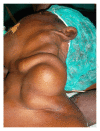
Figure 1
Figure 1
Anterolateral view of the cervical AVM.
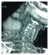
Figure 2
Figure 2
CT scan image of the cervical AVM. Anterolateral view showing connections with carotid, jugular, and subclavian.
Figure 3
Figure 3
CT scan. Posterior view of cervical AVM showing connections with vertebral.
Figure 4
Figure 4
Scan reconstruction showing the contrast filing the cervical AVM.
Figure 5
Figure 5
Preoperative view of the AVM exposed before surgical access.
Figure 6
Figure 6
Operative view of the external carotid artery connected to AVM.

References

    1. Lee B.-B., Do Y. S., Yakes W., Kim D. I., Mattassi R., Hyon W. S. Management of arteriovenous malformations: a multidisciplinary approach. Journal of Vascular Surgery. 2004;39(3):590–600. doi: 10.1016/j.jvs.2003.10.048. - DOI - PubMed
    1. Greenberg J. Spontaneous arteriovenous malformations in the cervical area. Journal of Neurology, Neurosurgery and Psychiatry. 1970;33(3):303–309. doi: 10.1136/jnnp.33.3.303. - DOI - PMC - PubMed
    1. González-García R., Rubio-Correa I., Moreno-García C. Massive glosso-cervical arteriovenous malformation: the rationale for a challenging surgical resection. Journal of Clinical and Experimental Dentistry. 2014;6(4):e456–e459. doi: 10.4317/jced.51608. - DOI - PMC - PubMed
    1. Kim J. Y., Kim D. I., Do Y. S., et al. Surgical treatment for congenital arteriovenous malformation: 10 years' experience. European Journal of Vascular and Endovascular Surgery. 2006;32(1):101–106. doi: 10.1016/j.ejvs.2006.01.004. - DOI - PubMed
    1. Lidsky M. E., Markovic J. N., Miller M. J., Jr., Shortell C. K. Analysis of the treatment of congenital vascular malformations using a multidisciplinary approach. Journal of Vascular Surgery. 2012;56(5):1355–1362. doi: 10.1016/j.jvs.2012.04.066. - DOI - PubMed

LinkOut - more resources