

Pathogenic Role of a Proliferation-Inducing Ligand (APRIL) in Murine IgA Nephropathy
- PMID: 26348210
- PMCID: PMC4562625
- DOI: 10.1371/journal.pone.0137044
Pathogenic Role of a Proliferation-Inducing Ligand (APRIL) in Murine IgA Nephropathy
Abstract
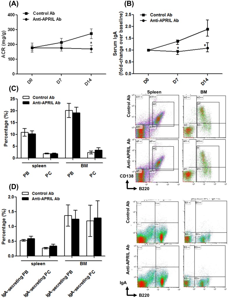
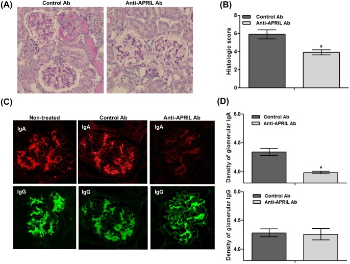
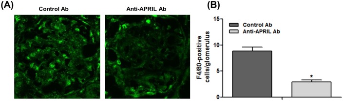
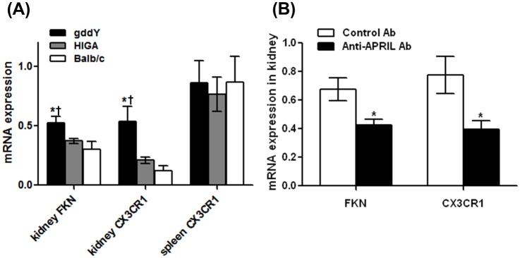
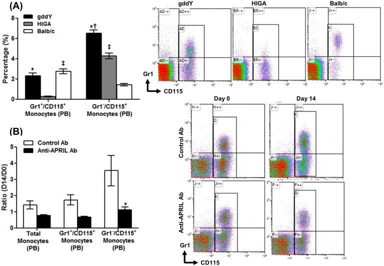
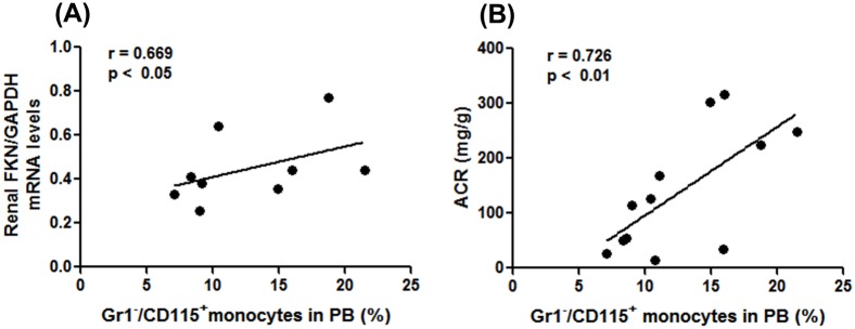
A proliferation-inducing ligand (APRIL) is a member of the tumor necrosis factor (TNF) superfamily. Despite advances in clinical and genetic studies, the details of the pathological roles of APRIL in IgA nephropathy (IgAN) remain to be fully defined. The present study aimed to further assess the pathological role of APRIL using a mouse model of IgAN. Mice with IgAN designated "grouped ddY" (gddY) were intraperitoneally administered an anti-APRIL monoclonal antibody (anti-APRIL Ab) or control IgG (Control Ab) twice each week for 2 weeks starting during the early stage of IgAN (6-7 weeks of age). Urinary albumin, serum IgA, and glomerular IgA deposition were evaluated. We further assessed the inflammatory responses during treatment by measuring the levels of the chemokine fractalkine (FKN) and its receptor CX3CR1 as well as the level of peripheral blood monocytosis. Anti-APRIL Ab treatment significantly decreased albuminuria and tissue damage combined with decreases in serum IgA levels and deposition of glomerular IgA. In contrast, the abundance of IgA+/B220+ or CD138+/B220+ B cells in the spleen and bone marrow, respectively, was unchanged. Treating gddY mice with anti-April Ab reduced the overexpression of FKN/CX3CR1 in the kidney and the increase in the population of circulating Gr1-/CD115+ monocytes. The size of the population of Gr1-/CD115+ monocytes correlated with renal FKN and urinary albumin levels. Moreover, mice treated with anti-APRIL Ab exhibited reduced progression of IgAN, serum IgA levels, and glomerular IgA deposition as well as an attenuated inflammatory process mediated by FKN-associated activation of monocytes. To the best of our knowledge, this is the first study to implicate the APRIL signal transduction pathway in the pathogenesis of nephrogenic IgA production. Moreover, our findings identify APRIL as a potential target of therapy.
Conflict of interest statement
Figures
Similar articles
-
A Proliferation Inducing Ligand (APRIL) targeted antibody is a safe and effective treatment of murine IgA nephropathy.Kidney Int. 2019 Jul;96(1):104-116. doi: 10.1016/j.kint.2019.01.031. Epub 2019 Mar 16. Kidney Int. 2019. PMID: 31027890
-
Crucial Role of AIM/CD5L in the Development of Glomerular Inflammation in IgA Nephropathy.J Am Soc Nephrol. 2020 Sep;31(9):2013-2024. doi: 10.1681/ASN.2019100987. Epub 2020 Jul 1. J Am Soc Nephrol. 2020. PMID: 32611589 Free PMC article.
-
Determination of severity of murine IgA nephropathy by glomerular complement activation by aberrantly glycosylated IgA and immune complexes.Am J Pathol. 2012 Oct;181(4):1338-47. doi: 10.1016/j.ajpath.2012.06.038. Epub 2012 Aug 5. Am J Pathol. 2012. PMID: 22871574 Free PMC article.
-
New Insights and Future Perspectives of APRIL in IgA Nephropathy.Int J Mol Sci. 2024 Sep 26;25(19):10340. doi: 10.3390/ijms251910340. Int J Mol Sci. 2024. PMID: 39408691 Free PMC article. Review.
-
Lessons from IgA Nephropathy Models.Int J Mol Sci. 2024 Oct 25;25(21):11484. doi: 10.3390/ijms252111484. Int J Mol Sci. 2024. PMID: 39519036 Free PMC article. Review.
Cited by
-
Safety, Tolerability, Pharmacokinetics, and Pharmacodynamics of VIS649 (Sibeprenlimab), an APRIL-Neutralizing IgG2 Monoclonal Antibody, in Healthy Volunteers.Kidney Int Rep. 2022 Feb 8;7(5):993-1003. doi: 10.1016/j.ekir.2022.01.1073. eCollection 2022 May. Kidney Int Rep. 2022. PMID: 35570983 Free PMC article.
-
BP180 Is Critical in the Autoimmunity of Bullous Pemphigoid.Front Immunol. 2017 Dec 8;8:1752. doi: 10.3389/fimmu.2017.01752. eCollection 2017. Front Immunol. 2017. PMID: 29276517 Free PMC article. Review.
-
Clinical and histological features and therapeutic strategies for IgA nephropathy.Clin Exp Nephrol. 2019 Sep;23(9):1089-1099. doi: 10.1007/s10157-019-01735-4. Epub 2019 Apr 9. Clin Exp Nephrol. 2019. PMID: 30968243 Review.
-
Perspectives on how mucosal immune responses, infections and gut microbiome shape IgA nephropathy and future therapies.Theranostics. 2020 Sep 15;10(25):11462-11478. doi: 10.7150/thno.49778. eCollection 2020. Theranostics. 2020. PMID: 33052226 Free PMC article. Review.
-
What is new in the pathogenesis and treatment of IgA glomerulonephritis.World J Nephrol. 2024 Dec 25;13(4):98709. doi: 10.5527/wjn.v13.i4.98709. World J Nephrol. 2024. PMID: 39723359 Free PMC article. Review.
References
-
- Ikezumi Y, Suzuki T, Imai N, Ueno M, Narita I, Kawachi H, et al. Histological differences in new-onset IgA nephropathy between children and adults. Nephrol Dial Transplant, 2006;21: 3466–3474. - PubMed
-
- Yoshikawa N, Iijima K, Maehara K, Yoshiara S, Yoshiya K, Matsuo T, et al. Mesangial changes in IgA nephropathy in children. Kidney Int. 1987;32: 585–589. - PubMed
-
- Li PK, Ho KK, Szeto CC, Yu L, Lai FM. Prognostic indicators of IgA nephropathy in the Chinese—clinical and pathological perspectives. Nephrol Dial Transplant. 2002;17: 64–69. - PubMed
Publication types
MeSH terms
Substances
LinkOut - more resources
Full Text Sources
Other Literature Sources
Molecular Biology Databases
Research Materials
Miscellaneous
