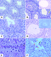
Histological changes of testes in growth hormone transgenic mice with high plasma level of GH and insulin-like growth factor-1
- PMID: 26348370
- PMCID: PMC4820830
- DOI: 10.5603/fhc.a2015.0024
Histological changes of testes in growth hormone transgenic mice with high plasma level of GH and insulin-like growth factor-1
Abstract
Introduction: Overexpression of growth hormone (GH) leads to increase in insulin-like growth factor-1 (IGF-1) plasma level, stimulation of growth and increase in body size, organomegaly and reduced body fat. The action of GH affects all the organs and transgenic mice that overexpress bovine GH (bGH mice) serve as convenient model to study somatotropic axis. Male mice overexpressing GH are fertile, however, they show reduced overall lifespan as well as reproductive life span. The aim of the study was to evaluate the morphology and expression of androgen receptor (AR) and luteinizing hormone receptor (LHR) of bGH mice testes.
Material and methods: The experiment was performed on 6 and 12 month-old bGH male mice and 6 and 12 month-old wild type (WT) littermates (8 animals in each group). The morphology of testes was evaluated on deparaffinized sections stained by the periodic acid-Schiff (PAS) method. Expression of AR and LHR was investigated by immunohistochemistry and diameters of seminiferous tubules (ST) were measured on round cross sections of ST.
Results: We noted larger testes in 6-month bGH mice as compared to normal WT littermates. The morpho-logical observations revealed essentially normal structure of Leydig cells, seminiferous epithelium and other morphological structures. However, some changes like tubules containing only Sertoli cells, tubules with arrested spermatogenesis or vacuoles in seminiferous epithelium could be attributed to the overexpression of GH. In contrast to WT mice, 12 month-old bGH mice displayed first symptoms of testicular aging. The immunoexpres-sion of AR and LHR was decreased in 12 month-old bGH males as compared to 12 month-old WT mice and younger animals.
Conclusion: Chronic exposure to elevated GH level accelerates testicular aging and thus potentially may change response of Leydig cells to LH and Sertoli and germ cells to testosterone.
Keywords: GH transgenic mice; IHC; LHR; androgen receptor; testis histology.
Figures


Similar articles
-
Growth response and androgen receptor expression in seminal vesicles from aging transgenic mice expressing human or bovine growth hormone genes.Endocrinology. 1992 Oct;131(4):2016-23. doi: 10.1210/endo.131.4.1396345. Endocrinology. 1992. PMID: 1396345
-
Fibroblast growth factor 21, fibroblast growth factor receptor 1, and β-Klotho expression in bovine growth hormone transgenic and growth hormone receptor knockout mice.Growth Horm IGF Res. 2016 Oct-Dec;30-31:22-30. doi: 10.1016/j.ghir.2016.08.003. Epub 2016 Aug 24. Growth Horm IGF Res. 2016. PMID: 27585733
-
Age-related alterations in pituitary and testicular functions in long-lived growth hormone receptor gene-disrupted mice.Endocrinology. 2007 Dec;148(12):6019-25. doi: 10.1210/en.2007-0837. Epub 2007 Sep 13. Endocrinology. 2007. PMID: 17872367
-
Neuroendocrine and reproductive consequences of overexpression of growth hormone in transgenic mice.Proc Soc Exp Biol Med. 1994 Sep;206(4):345-59. doi: 10.3181/00379727-206-43771. Proc Soc Exp Biol Med. 1994. PMID: 8073044 Review.
-
Effects of growth hormone overexpression and growth hormone resistance on neuroendocrine and reproductive functions in transgenic and knock-out mice.Proc Soc Exp Biol Med. 1999 Nov;222(2):113-23. doi: 10.1046/j.1525-1373.1999.d01-121.x. Proc Soc Exp Biol Med. 1999. PMID: 10564535 Review.
Cited by
-
Hallmarks of Testicular Aging: The Challenge of Anti-Inflammatory and Antioxidant Therapies Using Natural and/or Pharmacological Compounds to Improve the Physiopathological Status of the Aged Male Gonad.Cells. 2021 Nov 10;10(11):3114. doi: 10.3390/cells10113114. Cells. 2021. PMID: 34831334 Free PMC article. Review.
-
Excess Growth Hormone Triggers Inflammation-Associated Arthropathy, Subchondral Bone Loss, and Arthralgia.Am J Pathol. 2023 Jun;193(6):829-842. doi: 10.1016/j.ajpath.2023.02.010. Epub 2023 Mar 3. Am J Pathol. 2023. PMID: 36870529 Free PMC article.
-
Growth Hormone and Insulin-Like Growth Factor Action in Reproductive Tissues.Front Endocrinol (Lausanne). 2019 Nov 12;10:777. doi: 10.3389/fendo.2019.00777. eCollection 2019. Front Endocrinol (Lausanne). 2019. PMID: 31781044 Free PMC article. Review.
-
GH and ageing: Pitfalls and new insights.Best Pract Res Clin Endocrinol Metab. 2017 Feb;31(1):113-125. doi: 10.1016/j.beem.2017.02.005. Epub 2017 Feb 24. Best Pract Res Clin Endocrinol Metab. 2017. PMID: 28477727 Free PMC article. Review.
-
Evaluation of sex hormone profile and semen parameters in acromegalic male patients.J Endocrinol Invest. 2021 Dec;44(12):2799-2808. doi: 10.1007/s40618-021-01593-6. Epub 2021 May 28. J Endocrinol Invest. 2021. PMID: 34050506
References
-
- Berryman DE, List EO, Coschigano KT, et al. Comparing adiposity profiles in three mouse models with altered GH signaling. Growth Horm IGF Res. 2004;14:309–318. - PubMed
-
- Iida K, Del Rincon JP, Kim DS, et al. Tissue-specific regulation of growth hormone receptor and insulin like growth factor-I gene expression in the pituitary and liver of GH deficient (lit/lit) mice and transgenic mice that overexpress bovine GH (bGH) or a bGH antagonist. Endocrinology. 2004;145:1564–1570. - PubMed
MeSH terms
Substances
Grants and funding
LinkOut - more resources
Full Text Sources
Other Literature Sources
Research Materials
Miscellaneous

