

Comparing computer-generated and pathologist-generated tumour segmentations for immunohistochemical scoring of breast tissue microarrays
- PMID: 26348443
- PMCID: PMC4651129
- DOI: 10.1038/bjc.2015.309
Comparing computer-generated and pathologist-generated tumour segmentations for immunohistochemical scoring of breast tissue microarrays
Abstract
Background: Tissue microarrays (TMAs) have become a valuable resource for biomarker expression in translational research. Immunohistochemical (IHC) assessment of TMAs is the principal method for analysing large numbers of patient samples, but manual IHC assessment of TMAs remains a challenging and laborious task. With advances in image analysis, computer-generated analyses of TMAs have the potential to lessen the burden of expert pathologist review.
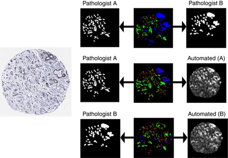
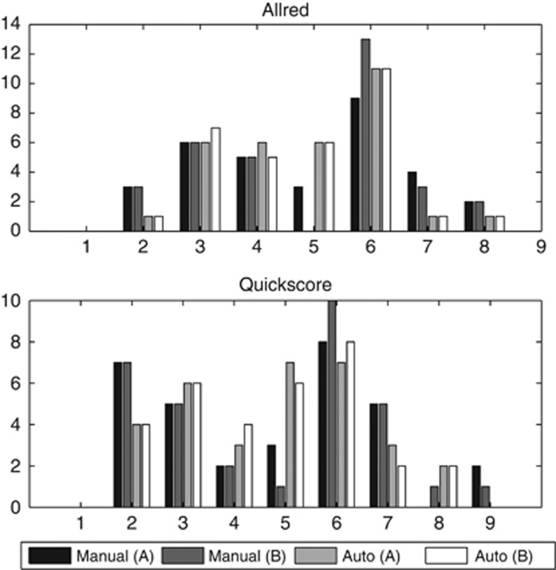
Methods: In current commercial software computerised oestrogen receptor (ER) scoring relies on tumour localisation in the form of hand-drawn annotations. In this study, tumour localisation for ER scoring was evaluated comparing computer-generated segmentation masks with those of two specialist breast pathologists. Automatically and manually obtained segmentation masks were used to obtain IHC scores for thirty-two ER-stained invasive breast cancer TMA samples using FDA-approved IHC scoring software.
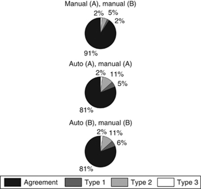
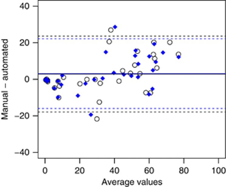
Results: Although pixel-level comparisons showed lower agreement between automated and manual segmentation masks (κ=0.81) than between pathologists' masks (κ=0.91), this had little impact on computed IHC scores (Allred; =0.91, Quickscore; =0.92).
Conclusions: The proposed automated system provides consistent measurements thus ensuring standardisation, and shows promise for increasing IHC analysis of nuclear staining in TMAs from large clinical trials.
Figures
References
-
- Allred DC, Bustamante MA, Daniel CO (1990) Immunocytochemical analysis of estrogen receptors in human breast carcinomas. Evaluation of 130 cases and review of the literature regarding concordance with bio- chemical assay and clinical relevance. Arch Surg 125(1): 107–113. - PubMed
-
- Arihiro K, Umemura S, Kurosumi M (2007) Comparison of evaluations for hormone receptors in breast carcinoma using two manual and three automated immunohistochemical assays. Am J Clin Pathol 127(3): 356–365. - PubMed
-
- Coates AS, Millar EKA, O'Toole SA, Molloy TJ, Viale G, Goldhirsch A, Regan MM, Gelber RD, Sun Z, Castiglione-Gertsch M, Gusterson B, Musgrove EA, Sutherland RL (2012) Prognostic interaction between expression of p53 and estrogen receptor in patients with node- negative breast cancer: results from IBCSG Trials VIII and IX. Breast Cancer Res 14: R143. - PMC - PubMed
-
- Cohen J (1968) Weighted kappa: nominal scale agreement with provision for scaled disagreement or partial credit. Psychol Bull 70(4): 213–220. - PubMed
Publication types
MeSH terms
Substances
Grants and funding
LinkOut - more resources
Full Text Sources
Other Literature Sources
Medical
Molecular Biology Databases
