. 2016 Feb;23(2):196-204.
doi: 10.1038/gt.2015.93. Epub 2015 Sep 8.

Photoreceptor rescue by an abbreviated human RPGR gene in a murine model of X-linked retinitis pigmentosa

Affiliations

Photoreceptor rescue by an abbreviated human RPGR gene in a murine model of X-linked retinitis pigmentosa

B S Pawlyk et al. Gene Ther. 2016 Feb.

Abstract

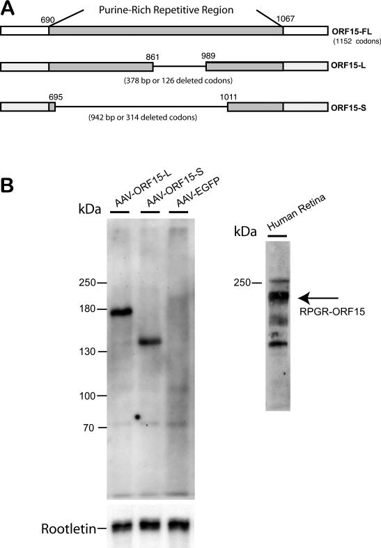
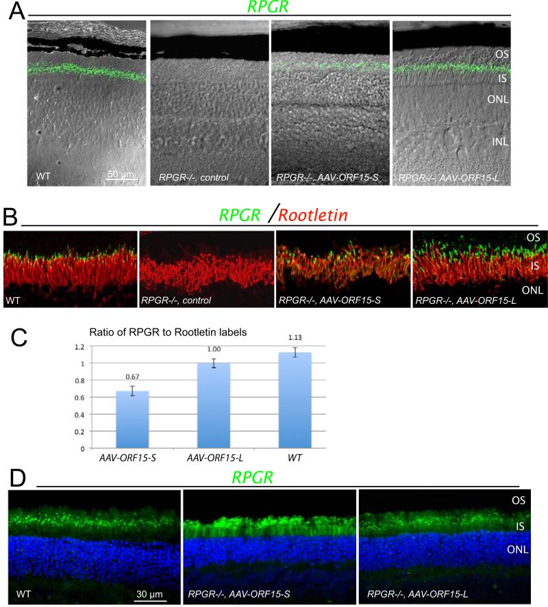
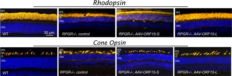
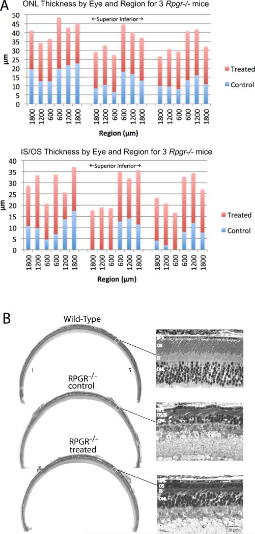
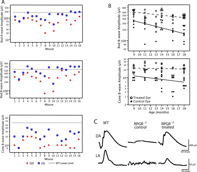
The X-linked RP3 gene codes for the ciliary protein RPGR and accounts for over 10% of inherited retinal degenerations. The critical RPGR-ORF15 splice variant contains a highly repetitive purine-rich linker region that renders it unstable and difficult to adapt for gene therapy. To test the hypothesis that the precise length of the linker region is not critical for function, we evaluated whether adeno-associated virus-mediated replacement gene therapy with a human ORF15 variant containing in-frame shortening of the linker region could reconstitute RPGR function in vivo. We delivered human RPGR-ORF15 replacement genes with deletion of most (314 codons, 'short form') or 1/3 (126 codons, 'long form') of the linker region to Rpgr null mice. Human RPGR-ORF15 expression was detected post treatment with both forms of ORF15 transgenes. However, only the long form correctly localized to the connecting cilia and led to significant functional and morphological rescue of rods and cones. Thus the highly repetitive region of RPGR is functionally important but that moderate shortening of its length, which confers the advantage of added stability, preserves its function. These findings provide a theoretical basis for optimizing replacement gene design in clinical trials for X-linked RP3.


Figures

Figure 1
Figure 1
(A) Maps of the native human RPGR ORF15 coding region and both shortened forms of AAV-delivered human ORF15cDNA. (B) Immunoblots for the two recombinant forms of human RPGR-ORF15. AAV delivery of the small-deletion human cDNA (AAV-ORF15-L, “long form”) leads to expression of a human RPGR-ORF15 protein of ~ 160 kD in size. AAV delivery of the large-deletion human cDNA (AAV-ORF15-S, “short form”) leads to expression of a protein of ~130 kD in size. Both forms of human RPGR-ORF15 protein are smaller than endogenous human RPGR ORF15 found in human retinal tissue (~ 200 kD).
Figure 2
Figure 2
RPGR ORF15 expression in Rpgr−/− mouse retinas following subretinal delivery of AAV-RPGR ORF15. (A) Fluorescence images of both the short (ORF15-S) and long (ORF15- L) forms of human RPGR ORF15 protein expression superimposed on Nomarski images to illustrate the layers of the outer retina. Staining of unfixed frozen retinal sections was performed at 3 weeks following treatment at 1-2 months of age. (B) Fluorescence images of both forms of human RPGR ORF15 co-localized with rootletin. Similar to WT, both forms of human RPGR ORF15 correctly localized to the photoreceptor connecting cilium just distal to rootletin. RPE, retinal pigment epithelium; OS, outer segment; CC (TZ), connecting cilium (transition zone); IS, inner segment; ONL, outer nuclear layer. (C) Ratio of hRPGR fluorescent particles to fluorescent rootletin fibers at the connecting cilium for Rpgr −/− eyes (n=3) treated with ORF15-S, Rpgr −/− eyes (n=3) treated with ORF15-L, and wt eyes (n=3). Counts were obtained for both rootletin within the inner segment and RPGR just distal to rootletin over a 100μm length of midperipheral retina. Values are means ± 1 standard error). (D) Expression pattern of short and long form ORF15 protein in fixed floating retinal sections of Rpgr−/− mice. Sections were stained for human RPGR ORF15 protein localization 4-6 weeks following treatment at 2-3 months of age. In wt retina, murine RPGR ORF15 protein is seen as a discrete green fluorescent signal (dots) occupying the region between the photoreceptor inner and outer segments, at the level of the transition zone or connecting cilium. In contrast, the fluorescent signal for the short form of ORF15 (AAV-ORF15-S) is not limited to level of the photoreceptor connecting cilium but is also seen as diffuse signal throughout the inner and outer segments as well. The fluorescent signal for the long form of ORF15 shows very little, if any mislocalization, and is largely limited to the connecting cilium region similar to wt. OS, outer segment; CC (TZ), connecting cilium (transition zone); IS, inner segment; ONL, outer nuclear layer.
Figure 3
Figure 3
Immunohistochemical (yellow) analyses of rod and cone photoreceptors in treated (short and long form of ORF15) and control Rpgr−/− mouse retinas at age 13 months (6-months post injection). In the Rpgr−/− mouse retina treated with the short form of ORF15 (AAV-ORF15-S), rhodopsin and cone opsin (mixed S & M cones in the inferior retina) mislocalization staining patterns are virtually indistinguishable from those seen in the control retina. Note the cone opsin mislocalization in the inner segments and synaptic layer in both of these mouse retinas.Similarly, rod and cone outer segments are shortened and disorganized with a reduced outer nuclear layer compared to an age matched wt retina. In contrast, in the Rpgr−/− mouse retina treated with the long form of ORF15 (AAV-ORF15-L) rhodopsin shows outer segment partitioning similar to WT mouse retina. Also in the ORF15 long form treated retina rod outer segments are longer and well organized and the ONL is thicker compared with the control retina. Cone opsin staining shows more numerous cone photoreceptors with elongated and well-organized outer segments in the ORF15 long form treated Rpgr−/− mouse retina compared with control.
Figure 4
Figure 4
Rescue of photoreceptor cells following treatment with RPGR ORF15-L in Rpgr−/− mice. (A) Shown are stacked bar graphs for ONL thickness (top) and IS/OS length (bottom) for treated (red) and fellow control (blue) eyes in 3 mice at 18 months of age. (B) Representative light micrographs from a WT mouse and an ORF15-L treated and fellow control eye from an Rpgr−/− mouse at 18 months of age. Images were taken from the mid periphery along the vertical meridian in the superior retina,
Figure 5
Figure 5
(A) Rod a-wave, rod b-wave, and cone b-wave amplitudes from 16 Rpgr−/− mice at 11-14 months of age following treatment with RPGR ORF15-L. Control eyes (OD) showed disproportionate loss of cone b-wave amplitude relative to rod b-wave amplitude compared with the lower limits for wild-type mice. Mean values for all three measures were significantly different between eyes (p<0.001). (B) Scatterplots of ERG amplitude for 22 Rpgr−/− mice between 9 and 18 months of age on a log scale for the dark-adapted (rod) b-wave (upper graph) and light-adapted (cone) b-wave (lower graph) following treatment with RPGR ORF15-L. Data points have been shifted slightly horizontally for each age group to minimize data overlap. The regression lines for treated and control eyes were fitted by repeated measures longitudinal regression using PROC Mixed of SAS based on all available data. (C) Representative dark-adapted (DA) and light-adapted (LA) ERG waveforms from a pair of ORF15-L treated and fellow control Rpgr−/− eyes at 18 months of age. WT (age-matched) ERG waveforms are shown for comparison. ERG a-wave and b-wave are labeled a and b, respectively.

References

    1. Bader I, Brandau O, Achatz H, Apfelstedt-Sylla E, Hergersberg M, Lorenz B, et al. X-linked retinitis pigmentosa: RPGR mutations in most families with definite X linkage andclustering of mutations in a short sequence stretch of exon ORF15. Invest Ophthalmol Vis Sci. 2003;44(4):1458–63. - PubMed
    1. Pelletier V, Jambou M, Delphin N, Zinovieva E, Stum M, Gigarel N, et al. Comprehensive survey of mutations in RP2 and RPGR in patients affected with distinctretinal dystrophies: genotype-phenotype correlations and impact on genetic counseling. Hum Mutat. 2007;28(1):81–91. - PubMed
    1. Branham K, Othman M, Brumm M, Karoukis AJ, Atmaca-Sonmez P, Yashar BM, et al. Mutations in RPGR and RP2 Account for 15% of Males with Simplex RetinalDegenerative Disease. Invest Ophthalmol Vis Sci. 2012;53(13):8232–7. - PMC - PubMed
    1. Churchill JD, Bowne SJ, Sullivan LS, Lewis RA, Wheaton DK, Birch DG, et al. Mutations in the X-linked retinitis pigmentosa genes RPGR and RP2 found in 8.5% offamilies with a provisional diagnosis of autosomal dominant retinitis pigmentosa. Invest Ophthalmol Vis Sci. 2013;54(2):1411–6. - PMC - PubMed
    1. Hong DH, Pawlyk BS, Shang J, Sandberg MA, Berson EL, Li T. A retinitis pigmentosaGTPase regulator (RPGR)-deficient mouse model for X-linked retinitis pigmentosa(RP3). Proc Natl Acad Sci USA. 2000;97(7):3649–54. - PMC - PubMed

Publication types