. 2015 Sep 8;11(9):e1005396.
doi: 10.1371/journal.pgen.1005396. eCollection 2015 Sep.

The Chromatin Protein DUET/MMD1 Controls Expression of the Meiotic Gene TDM1 during Male Meiosis in Arabidopsis

Affiliations

The Chromatin Protein DUET/MMD1 Controls Expression of the Meiotic Gene TDM1 during Male Meiosis in Arabidopsis

Sébastien Andreuzza et al. PLoS Genet. .

Abstract

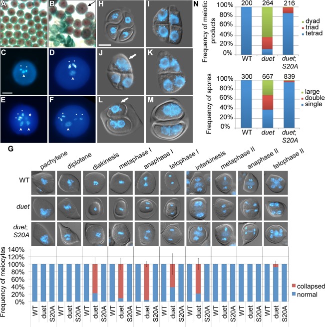
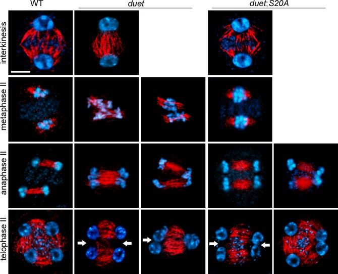
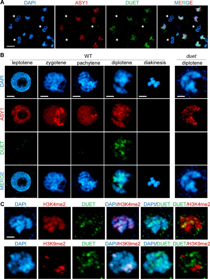
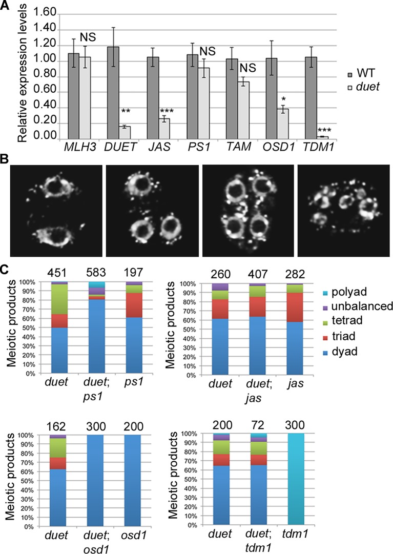
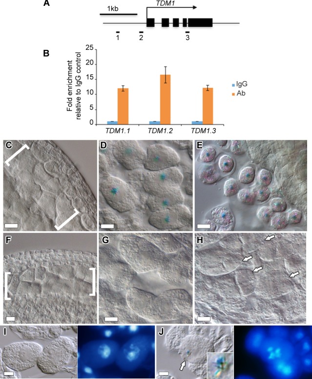
Meiosis produces haploid cells essential for sexual reproduction. In yeast, entry into meiosis activates transcription factors which trigger a transcriptional cascade that results in sequential co-expression of early, middle and late meiotic genes. However, these factors are not conserved, and the factors and regulatory mechanisms that ensure proper meiotic gene expression in multicellular eukaryotes are poorly understood. Here, we report that DUET/MMD1, a PHD finger protein essential for Arabidopsis male meiosis, functions as a transcriptional regulator in plant meiosis. We find that DUET-PHD binds H3K4me2 in vitro, and show that this interaction is critical for function during meiosis. We also show that DUET is required for proper microtubule organization during meiosis II, independently of its function in meiosis I. Remarkably, DUET protein shows stage-specific expression, confined to diplotene. We identify two genes TDM1 and JAS with critical functions in cell cycle transitions and spindle organization in male meiosis, as DUET targets, with TDM1 being a direct target. Thus, DUET is required to regulate microtubule organization and cell cycle transitions during male meiosis, and functions as a direct transcription activator of the meiotic gene TDM1. Expression profiling showed reduced expression of a subset comprising about 12% of a known set of meiosis preferred genes in the duet mutant. Our results reveal the action of DUET as a transcriptional regulator during male meiosis in plants, and suggest that transcription of meiotic genes is under stagewise control in plants as in yeast.


Conflict of interest statement

The authors have declared that no competing interests exist.

Figures

Fig 1
Fig 1. DUET PHD is a reader of H3K4me2.
(A) Schematic representation of DUET protein. DUET encodes a 704 amino acid plant specific protein with a nuclear localization sequence (NLS), a region showing homology with the meiotic gene SWITCH1 (SWI1), and a C-terminal Plant Homeo Domain (PHD). (B) Calf thymus histone pull down followed by western blot with the indicated antibodies. BPTF PHD2 is used as positive control. (C) Peptide pull downs followed by western blot with anti-GST antibody. (D) The PHD finger of DUET is conserved across eukaryotes. Sequence alignment of DUET PHD finger with characterized homologues showing highest homology obtained by PSI-BLAST. DUET PHD shares 46% identity with human MLL5 PHD (E = 9e-08), 44% identity with S.cerevisiae Set3 PHD (E = 2e-07), and 41% identity with human BPTF PHD2 (E = 3e-05). Conserved and similar residues are in red and blue respectively, conserved C4HC3 structural residues are in bold green. White circles indicate mutation sites in the triple mutant (C21A/C24A/H29A), and black circles indicate the point mutations S20A and W27A. (E) Histone peptide pull downs with methylated H3K4 peptides and the indicated mutant DUET PHD fingers. (F) Representative Alexander staining of T1 duet plants transformed with a wild-type DUET construct (WT). Left panel: fully viable pollen (purple) indicating full rescue; middle panel: mix of viable and dead pollen (green) indicating partial rescue; right panel: absence of pollen indicating no rescue. (G) Quantification of rescue phenotypes obtained for independent T1 duet plants transformed with the indicated construct. WT, WT full length DUET construct; n, number of independent transformants analyzed per construct.
Fig 2
Fig 2. Partial complementation of duet reveals a role in chromosome segregation and cytokinesis.
(A and B) Alexander staining of mature anthers of WT (A) and partially complemented duet with DUET-S20A (duet;S20A) (B). Viable pollen is purple; dead pollen is green; arrow points to an enlarged pollen grain. (C) DAPI stained WT pollen with a male germ unit (MGU) comprising two condensed sperm nuclei (white dots) and one decondensed vegetative cell nucleus (arrowhead).(D-F) DAPI stained enlarged pollen from duet;S20A; scale bar = 10 μm. (D) DAPI stained large duet;S20A pollen grain with a normal MGU. (E) DAPI stained large duet;S20A pollen grain with two MGUs; star indicates a condensed sperm-like cell out of focus. (F) DAPI stained large duet;S20A pollen grain with four sperm-like condensed cells, and one decondensed vegetative-like cell. (G) Analysis of collapse during meiosis in WT, duet and duet;S20A. Top panel: analysis of cytoplasmic collapse during meiosis in the indicated backgrounds. Pictures are merged DIC and DAPI, which stains chromosomes blue. Bottom graph: stage-wise quantification of collapsed meiocytes. Data are presented as mean ± SD for at least 20 meiocytes per stage. (H-M) Microscopic analysis of tetrads from WT (H), duet (I-K) and duet;S20A (L and M). scale bar = 10 μm.
Fig 3
Fig 3. DUET is required for proper spindle organization during meiosis II.
Immunostaining of α-tubulin on male meiotic squashes from WT, duet, and duet;S20A. The meiotic stages are indicated for each row. Chromosomes were stained with DAPI (blue) tubulin, red. White arrow: absence of radial microtubule arrays (RMA) between nuclei; scale bar = 10 μm. At least 20 meiocytes were analyzed per stage and per genotype.
Fig 4
Fig 4. DUET is expressed during diplotene and localizes on euchromatin.
(A and B) Dual immunostaining of DUET (green) and ASY1 (red). (A) DUET is only expressed in ASY1 positive cells (white arrows), and not in other anther cells, including multinucleated tapetal cells (stars). Scale bar = 20 μm. (B) Detailed analysis of DUET expression during prophase stages, according to chromosome morphology and ASY1 pattern. Scale bar = 5 μm. (C) Dual immunostaining of DUET (green), and H3K4me2 (red, top panel), or H3K9me2 (red, bottom panel) chromatin marks. In each row, the first three pictures show single channels, the last three pictures are merged images of two channels. Scale bar = 5 μm.
Fig 5
Fig 5. DUET is required for proper expression of JAS and TDM1 during meiosis.
(A) Quantitative RT-PCR (qPCR) analysis of meiotic gene expression in WT and duet from anthers dissected from 0.5–0.6mm buds normalized on ACT11 expression. Data are presented as the mean ± SEM (error bars) of at least two independent experiments with at least three samples each. Statistically significant differences: P < 0.05 = (*); P < 0.01 = (**); P < 0.001 = (***); ns = no significant difference (t-test). (B) Representative images of major classes of male meiotic products obtained in F2 double mutant combinations. Chromosomes were stained with DAPI (white). Left to right: meiotic products with two, three, four and more than four nuclei respectively. (C) Quantification of male meiotic products described in (B), in the indicated genetic backgrounds.
Fig 6
Fig 6. TDM1 is a direct target of DUET.
(A) Schematic diagram of TDM1, representing the regions analyzed by ChIP-qPCR (black rectangles). Black boxes represent exons, the arrow represent the direction of transcription. (B) Quantification of ChIP relative to the IgG control by qPCR. Columns represent the mean, and error bars represent the standard error of the mean from 3 independent biological samples. (C-E) Expression of pTDM1::NLS-GUS during WT meiosis in whole mount anthers. (C) Early prophase, (D) late prophase, meiocytes are individualized by a callose envelope, (E) tetrad stage. (F-H) Expression of pTDM1::NLS-GUS during duet meiosis in whole mount anthers. (F) Early prophase, (G) late prophase, (H) tetrads. Brackets in (C) and (F) delineate meiocytes are arrows in (H) point to individual tetrads. All panels, scale bars 10 μm, except (D) and (G), 5 μm. (I,J) GUS staining (left panel), followed by DAPI staining of squashed anthers (right panel), allows staging of meiocytes based on chromosome morphology. (I) pachytene, (J) diakinesis. The arrow in (J) points to the GUS signal shown in the inset.

References

    1. Winter E. The Sum1/Ndt80 Transcriptional Switch and Commitment to Meiosis in Saccharomyces cerevisiae. Microbiol Mol Biol Rev. 2012;76: 1–15. 10.1128/MMBR.05010-11 - DOI - PMC - PubMed
    1. Werven FJ van, Amon A. Regulation of entry into gametogenesis. Philos Trans R Soc B Biol Sci. 2011;366: 3521–3531. 10.1098/rstb.2011.0081 - DOI - PMC - PubMed
    1. Bolcun-Filas E, Bannister LA, Barash A, Schimenti KJ, Hartford SA, Eppig JJ, et al. A-MYB (MYBL1) transcription factor is a master regulator of male meiosis. Development. 2011;138: 3319–3330. 10.1242/dev.067645 - DOI - PMC - PubMed
    1. Hayashi K, Yoshida K, Matsui Y. A histone H3 methyltransferase controls epigenetic events required for meiotic prophase. Nature. 2005;438: 374–378. 10.1038/nature04112 - DOI - PubMed
    1. White-Cooper H. Molecular mechanisms of gene regulation during Drosophila spermatogenesis. Reproduction. 2010;139: 11–21. 10.1530/REP-09-0083 - DOI - PubMed

Publication types