Comparative Study
. 2015 Oct 1;125(10):3891-903.
doi: 10.1172/JCI77995. Epub 2015 Sep 8.

Hepatocellular carcinoma originates from hepatocytes and not from the progenitor/biliary compartment

Comparative Study

Hepatocellular carcinoma originates from hepatocytes and not from the progenitor/biliary compartment

Xueru Mu et al. J Clin Invest. .

Abstract

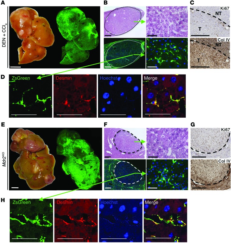
In many organs, including the intestine and skin, cancers originate from cells of the stem or progenitor compartment. Despite its nomenclature, the cellular origin of hepatocellular carcinoma (HCC) remains elusive. In contrast to most organs, the liver lacks a defined stem cell population for organ maintenance. Previous studies suggest that both hepatocytes and facultative progenitor cells within the biliary compartment are capable of generating HCC. As HCCs with a progenitor signature carry a worse prognosis, understanding the origin of HCC is of clinical relevance. Here, we used complementary fate-tracing approaches to label the progenitor/biliary compartment and hepatocytes in murine hepatocarcinogenesis. In genotoxic and genetic models, HCCs arose exclusively from hepatocytes but never from the progenitor/biliary compartment. Cytokeratin 19-, A6- and α-fetoprotein-positive cells within tumors were hepatocyte derived. In summary, hepatocytes represent the cell of origin for HCC in mice, and a progenitor signature does not reflect progenitor origin, but dedifferentiation of hepatocyte-derived tumor cells.


Figures

Figure 6
Figure 6. A6-, K19-, and AFP-positive liver progenitors within HCCs are derived from hepatocytes.
(AD) To determine the cellular origin of liver progenitor cells within HCCs, colocalization of the progenitor markers A6 (A), K19 (B), and AFP (C) with mGFP was determined by confocal microcopy in tumor and nontumor areas of mTom-mGFP mice whose hepatocytes had been labeled via AAV8-Tbg-Cre and that were subsequently treated with DEN and CCl4. Quantification of mGFP of A6-, K19-, and AFP-positive cells in tumor and nontumor areas (n = 10 each) (D). (EH) Colocalization of the progenitor/hepatoblast markers A6 (E, serial sections), K19 (F, fluorescence), and AFP (G, serial sections) with YFP in tumor and nontumor areas of Opn-CreERT2 mice treated with 25 injections of DEN or with DEN+CCl4. Quantification of YFP with A6-, K19-, or AFP-positive cells (H) in tumor or nontumor areas. **P < 0.01, ***P < 0.001 by 2-sided Student’s t test. ND, nondetectable. Scale bars: 50 μm (AC and insert in E); 100 μm (EG).
Figure 5
Figure 5. HCCs are not derived from HSCs.
(AD) Lrat-Cre Tg mice expressing the ZsGreen Cre reporter (n = 4) were treated with DEN and 20 injections of CCl4 for HCC induction. Representative images and fluorescent images of livers from DEN+CCl4-treated mice (A) as well as H&E- and Hoechst-stained frozen liver sections at low and high magnification show green fluorescent HSCs but no green fluorescent tumor cells derived from HSCs (B). Typical HCC features were confirmed by collagen IV staining and increased Ki67 expression (C). ZsGreen-positive cells colocalized with the HSC marker desmin (D). (EH) Mdr2KO mice expressing Lrat-Cre and the ZsGreen Cre reporter (n = 8) were sacrificed at 12 months of age. Representative images and fluorescent images of whole livers (E) as well as H&E- and Hoechst-stained frozen liver sections at low and high magnification show green fluorescent HSCs but no green fluorescent tumor cells derived from HSCs (F). Typical HCC features were confirmed by increased Ki67 expression and altered collagen IV staining (G). ZsGreen-positive cells colocalized with the HSC marker desmin (H). Scale bars: 5 mm (A and E); 500 μm (B, left panel and F, left panel); 50 μm (B, right panel and F, right panel); 500 μm (C and G); 50 μm (D and H).
Figure 4
Figure 4. HCCs derive from hepatocytes in the Mdr2KO HCC model.
Mdr2KO mice expressing the ZsGreen Cre reporter (n = 9) were infected with AAV8-Tbg-Cre to selectively label hepatocytes and sacrificed at 12 to 14 months of age. (AC) Representative images and fluorescent images of whole livers from Mdr2KO mice (A) as well as H&E- and Hoechst-stained frozen liver sections at low (B) and high (C) power, including a ZsGreen negative control. (D) Quantification of GFP-labeled hepatocytes and tumors (average of all mice). (E) Costaining demonstrated that HNF4α-positive tumor cells were ZsGreen positive. (F) Typical HCC features were confirmed by increased Ki67 expression and altered collagen IV staining. (G and H) HCC markers (G) and progenitor markers (H) were determined by qPCR (n = 5 control livers and n = 14 tumors). Scale bars: 1 cm (A); 300 μm (B); 50 μm (C and E); 500 μm (F). **P < 0.01 by Mann-Whitney U test.
Figure 3
Figure 3. Genotoxic HCC derives from hepatocytes.
mTom-mGFP Cre reporter mice (n = 10) were infected with AAV8-Tbg-Cre to selectively label hepatocytes, followed by treatment with DEN and 20 injections of CCl4 for HCC induction. (AC) Representative images and fluorescent images of livers from DEN+CCl4-treated mice (A) and H&E- and GFP-stained liver sections at low (B) and high (C) magnification, including an mTom-mGFP negative control. (D) Quantification of GFP-labeled hepatocytes and tumors (average of all mice, n = 10). (E) Typical HCC features were confirmed by collagen IV staining, increased Ki67 and glutamine synthetase levels, and altered Gp73 and β-catenin (β-Cat) staining. (F and G) HCC markers (F) and progenitor markers (G) were determined by qPCR. Scale bars: 1 cm (A); 1 mm (B); 50 μm (C); 300 μm (E). *P < 0.05, **P < 0.01, ***P < 0.01 by Kruskal-Wallis and Dunn’s post-hoc test.
Figure 2
Figure 2. HCC does not derive from the LPC/biliary compartment in the DEN+CCl4 model.
Tamoxifen-treated Opn-CreERT2 mice were treated with DEN followed by 20 injections (20×) of CCl4 (n = 6). (A and B) Representative macroscopic (A) and histological images (B) showing typical HCC features such as high Ki67 expression levels and disruption of the collagen IV meshwork. Col IV, collagen IV. (C and D) The cholangiocyte/LPC compartment was YFP positive, but no HCCs were YFP positive (n = 6). (E) HCC and progenitor/hepatoblast markers determined by qPCR. (FI) mTom-mGFP Cre reporter mice (n = 5) were treated with tamoxifen, followed by administration of DEN and 20 injections of CCl4 for HCC induction. Representative images and fluorescent images of livers (F) and H&E- and GFP-stained liver sections at low (G) and high (H) magnification, demonstrating mGFP-positive ducts in nontumor areas but not in tumor areas. Quantification of GFP-labeled K19-positive cholangiocytes and tumors (n = 5) (I). Sac, sacrifice. Scale bars: 1 cm (A and F); 100 μm (B and G); 50 μm (C and H). **P < 0.01, ***P < 0.001 by Mann-Whitney U test.
Figure 1
Figure 1. HCC does not derive from the LPC/biliary compartment.
Tamoxifen-treated Opn-CreERT2 mice expressing the YFP Cre reporter were subjected to 25 injections of DEN (n = 33). (A and B) Mice developed macroscopically (A) and microscopically (B) visible HCCs. Tam, tamoxifen. (C and D) While the K19-positive biliary/LPC compartment was tagged efficiently by YFP, no HCCs were YFP positive. HCCs were delineated by an α-SMA–positive border and infiltrated by myofibroblasts; HCCs displayed a disrupted reticulin meshwork, high Ki67 expression levels, focally expressed OPN and AFP, and were surrounded by a patchy K19-positive and YFP-positive DR. Scale bars: 1 cm (A and B); 100 μm (D). Chol, cholangiocyte; NT, nontumor; T, tumor.

Similar articles

Cited by

References

    1. Parkin DM, Bray F, Ferlay J, Pisani P. Estimating the world cancer burden: Globocan 2000. Int J Cancer. 2001;94(2):153–156. doi: 10.1002/ijc.1440. - DOI - PubMed
    1. Forner A, Llovet JM, Bruix J. Hepatocellular carcinoma. Lancet. 2012;379(9822):1245–1255. doi: 10.1016/S0140-6736(11)61347-0. - DOI - PubMed
    1. Miyajima A, Tanaka M, Itoh T. Stem/progenitor cells in liver development, homeostasis, regeneration, and reprogramming. Cell Stem Cell. 2014;14(5):561–574. doi: 10.1016/j.stem.2014.04.010. - DOI - PubMed
    1. Donati G, Watt FM. Stem cell heterogeneity and plasticity in epithelia. Cell Stem Cell. 2015;16(5):465–476. doi: 10.1016/j.stem.2015.04.014. - DOI - PubMed
    1. Clevers H. The intestinal crypt, a prototype stem cell compartment. Cell. 2013;154(2):274–284. doi: 10.1016/j.cell.2013.07.004. - DOI - PubMed

Publication types

MeSH terms