. 2015 Sep 8:5:13798.
doi: 10.1038/srep13798.

The identification of an integral membrane, cytochrome c urate oxidase completes the catalytic repertoire of a therapeutic enzyme

Affiliations

The identification of an integral membrane, cytochrome c urate oxidase completes the catalytic repertoire of a therapeutic enzyme

Nicola Doniselli et al. Sci Rep. .

Abstract

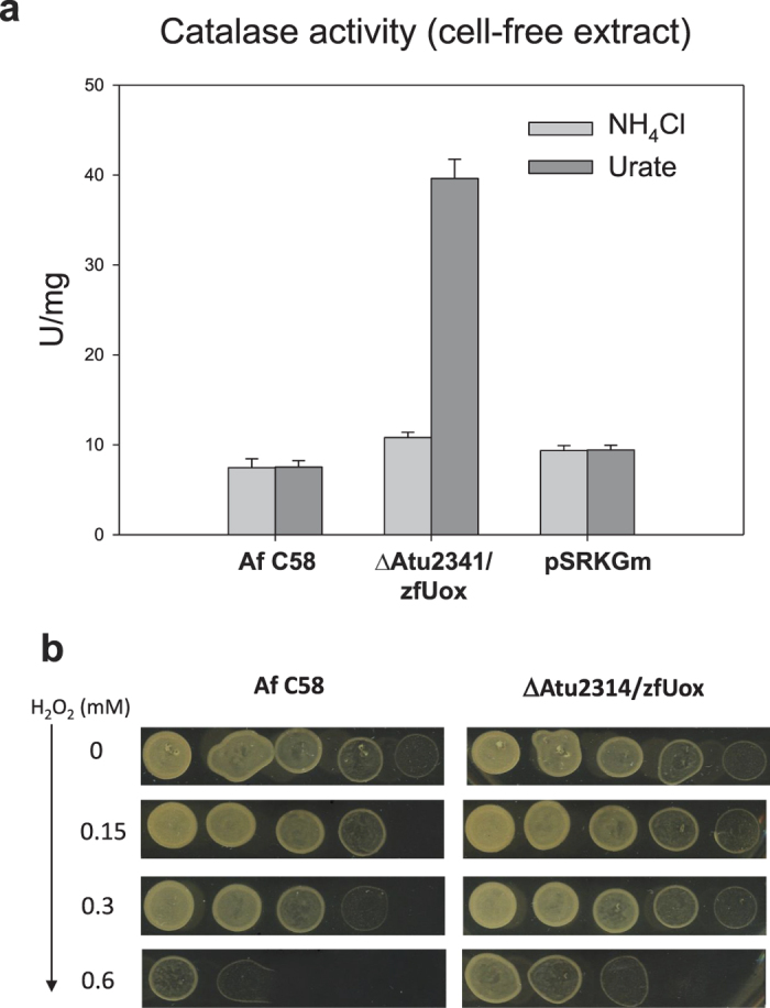
In living organisms, the conversion of urate into allantoin requires three consecutive enzymes. The pathway was lost in hominid, predisposing humans to hyperuricemia and gout. Among other species, the genomic distribution of the two last enzymes of the pathway is wider than that of urate oxidase (Uox), suggesting the presence of unknown genes encoding Uox. Here we combine gene network analysis with association rule learning to identify the missing urate oxidase. In contrast with the known soluble Uox, the identified gene (puuD) encodes a membrane protein with a C-terminal cytochrome c. The 8-helix transmembrane domain corresponds to DUF989, a family without similarity to known proteins. Gene deletion in a PuuD-encoding organism (Agrobacterium fabrum) abolished urate degradation capacity; the phenotype was fully restored by complementation with a cytosolic Uox from zebrafish. Consistent with H2O2 production by zfUox, urate oxidation in the complemented strain caused a four-fold increase of catalase. No increase was observed in the wild-type, suggesting that urate oxidation by PuuD proceeds through cytochrome c-mediated electron transfer. These findings identify a missing link in purine catabolism, assign a biochemical activity to a domain of unknown function (DUF989), and complete the catalytic repertoire of an enzyme useful for human therapy.


Figures

Figure 1
Figure 1. Identification of COG3748 as urate oxidase.
(a) Pathway for the conversion of urate into allantoin. (b) String association network obtained with COG3648, COG2351, and COG3195. Nodes represent gene families according to the COG classification. Edges represent predicted functional associations; stronger associations are shown as thicker lines. The node identified as a candidate urate oxidase (COG3748) is indicated with an arrow. (c) Map of urate oxidation capacity in complete genomes. The tree represents 431 distinct species possessing either the uox, hpxO, hpyO, or COG3748 genes or both the urah and urad genes. The presence (red) or the absence (blue) of genes in complete genomes is shown alongside the organism tree. Main taxonomic groups and organisms discussed in the text are indicated.
Figure 2
Figure 2. Experimental evidence for the urate oxidase activity of Atu2314 (COG3748).
Growth curves of wild-type and engineered A. fabrum C58 strains in M9 minimal medium supplemented with (a) urate or (b) ammonia as nitrogen source. (c) Urate utilization by concentrated cell cultures. Error bars represent standard deviations obtained from three independent experiments. (d) Uox activity of 250 μg of cell-free extracts as monitored by the decrease in absorbance at 293 nm; extracts were added (arrows) to 0.1 ml solutions containing 0.11 mM urate.
Figure 3
Figure 3. Sequence and structure organization of PuuD proteins.
(a) Multiple alignment of representative PuuD sequences from Agrobacterium fabrum (Af), Paracoccus denitrificans (Pd), Pseudomonas aeruginosa (Pa), Acidovorax avenae (Aa), and Ralstonia solanacearum (Rs). Accession numbers are reported in Supplementary Table S1. Colour shading of conserved residues is based on the larger alignment shown in Supplementary Fig. S7. The identified transmembrane helices in AfPuuD are indicated by gray bars and numbered with roman numerals. Blue arrowheads indicate the residues supposedly involved in heme coordination. The secondary structure elements of the cytochrome c domain are depicted above the alignment based on the comparison with the PDB structure 2d0w; helices are named according to the mitochondrial cytochrome c notation. (b) Membrane topology of AfPuuD. Residues are colored according to the Consurf analysis of the PuuD multiple alignment of Supplementary Fig. S7. Topology plots were obtained with the Protter program.
Figure 4
Figure 4. Catalase activity of PuuD and zfUox encoding strains grown on ammonia or urate.
(a) Catalase activity (U/mg) of free-cell extracts obtained by sonication of urate- and ammonia-grown cells collected during the exponential growth phase; pSRKGm designates Af C58 cells transformed with the empty vector. Error bars represent standard deviation obtained from three independent experiments. (b) Urate-grown strains plated on LB agar supplemented with increasing concentrations of hydrogen peroxide. Cells were collected during exponential growth phase, diluted at OD600 = 0.1 and spotted at progressive dilutions (1, 1:5, 1:25, 1:125, 1:625). Scanned plate images were adjusted with −40% luminosity and +40% contrast.
Figure 5
Figure 5. Distribution of urate oxidation genes in monoderm and diderm prokaryotes.
The occurrence of the different genes for urate oxidation in 341 prokaryotic species (see Fig. 1c) classified based on the presence of a single (monoderm) or a double (diderm) cell membrane.

References

    1. Terkeltaub R. Update on gout: new therapeutic strategies and options. Nat. Rev. Rheumatol. 6, 30–38 (2010). - PubMed
    1. Cheuk D. K. L., Chiang A. K. S., Chan G. C. F. & Ha S. Y. Urate oxidase for the prevention and treatment of tumour lysis syndrome in children with cancer. Cochrane Database Syst Rev. 8, CD006945 (2014). - PubMed
    1. Roche A. et al. Efficacy of Rasburicase in Hyperuricemia Secondary to Lesch-Nyhan Syndrome. Am. J. Kidney Dis. 53, 677–680 (2009). - PubMed
    1. Kratzer J. T. et al. Evolutionary history and metabolic insights of ancient mammalian uricases. Proc. Natl. Acad. Sci. USA 111, 3763–8 (2014). - PMC - PubMed
    1. Ramazzina I., Folli C., Secchi A., Berni R. & Percudani R. Completing the uric acid degradation pathway through phylogenetic comparison of whole genomes. Nat. Chem. Biol. 2, 144–148 (2006). - PubMed

Publication types

MeSH terms

LinkOut - more resources